Rebuilding method of real-time three-dimensional medical ultrasonic image

An ultrasonic image, real-time 3D technology, applied in 3D image processing, medical science, image data processing, etc., can solve problems such as deformation, inability to truly reflect the measured object, and shape distortion

Active Publication Date: 2010-09-01
SHANTOU INST OF UITRASONIC INSTR CO LTD
View PDF0 Cites 0 Cited by
  • Summary
  • Abstract
  • Description
  • Claims
  • Application Information

AI Technical Summary

Problems solved by technology

However, the volume image data obtained by the image processing device from the ultrasonic three-dimensional sensor is actually cube data, that is, the volume image data obtained from the ultrasonic three-dimensional sensor consists of a series of parallel two-dimensional slices, and these

Method used


Image

  • Rebuilding method of real-time three-dimensional medical ultrasonic image
  • Rebuilding method of real-time three-dimensional medical ultrasonic image
  • Rebuilding method of real-time three-dimensional medical ultrasonic image

Examples


Embodiment Construction

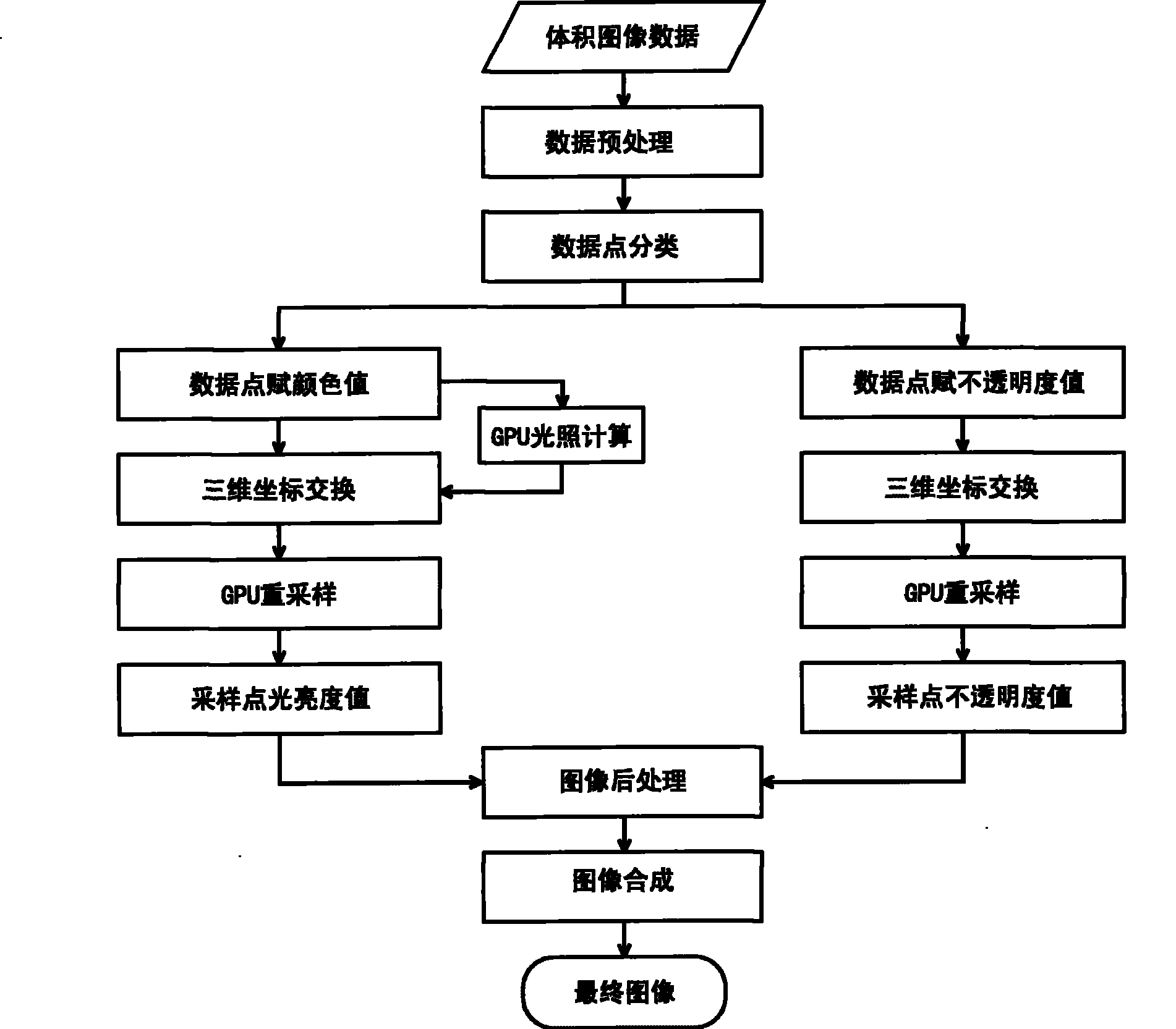
[0068] Such as figure 2 As shown, the reconstruction method of this real-time three-dimensional medical ultrasound image includes the following steps in sequence:

[0069] (1) Preprocessing the volume image data from the ultrasonic three-dimensional sensor to obtain the volume image data required for image reconstruction; this step can be performed in the central processing unit CPU.

[0070] After the volumetric image data from the ultrasonic three-dimensional sensor is obtained, the volumetric image data is firstly preprocessed, including format conversion of the original volumetric image data, elimination of redundant data, and export of the required volumetric image data.

[0071] The data obtained by the ultrasonic three-dimensional sensor is generally 16 bits or more, but the screen can only display 8-bit grayscale images, which requires us to convert the format of the original volume image data (convert the data of 16 bits or more to 8-bit data), so that it meets the ...


PUM


Abstract

The present invention provides a real time three-dimensional medical ultrasonic image reconstruction method, including steps in sequence: (1) preprocessing volume image data from an ultrasonic three-dimensional sensor to acquire required volume image data for reconstructing image; (2) classifying data points of the volume image data acquired in step (1), wherein data points of same material are classified into one classification; (3) executing value assignment to the data points, wherein the value assignment includes color value assignment and opacity coefficient value assignment to the data points; (4) executing three-dimensional transformation of coordinates of the volume image data in an image processor, wherein hexahedron data are converted into hyperboloid data; (5) resampling in theimage processor and converting original coordinate of data points into coordinate of data points in hyperboloid data; (6) synthesizing image. According to the invention, not only original image data are not lost, but also real-time display of image can be realized, thereby realizing real time reconstruction of three-dimensional medical ultrasonic image, and enabling finally formed image to truly reflect true forms of the tested objects.

Description

technical field [0001] The invention relates to image data processing, in particular to a method for reconstructing real-time three-dimensional medical ultrasonic images. Background technique [0002] The visualization and interpretation of traditional medical two-dimensional images has been widely accepted, but after the invention of volume image technology, since each volume image data (ie three-dimensional image data) contains a series of two-dimensional slices (ie two-dimensional image data) , the number of 2D slices that must be viewed is exploding, creating an urgent need to more intuitively observe volumetric image data. [0003] The main purpose of 3D visualization is to use computer graphics technology to construct a 3D model of an organ from a series of 2D slices obtained by medical imaging equipment (this series of 2D slices constitute volume image data), and it is very realistic. is displayed to the viewer, so that the viewer can intuitively observe the volumetr...

Claims


Application Information

Patent Timeline
IPC IPC(8): A61B8/00G06T15/00
Inventor 郭境峰王海潮蔡泽杭
Owner SHANTOU INST OF UITRASONIC INSTR CO LTD
Why Eureka
Try Eureka