Radiographic apparatus

A diagnostic device and X-ray technology, applied in the direction of diagnosis, clinical application of radiodiagnosis, and equipment for radiodiagnosis, etc., can solve problems such as reduction and increase in line dose, and achieve good depiction ability, reduced exposure dose, and good guidance. The effect of silk manipulation

CN101484073BInactive Publication Date: 2011-04-13SHIMADZU SEISAKUSHO CO LTD
1 Cites 0 Cited by

Patent Information

Authority / Receiving Office
CN · China
Patent Type
Patents(China)
Current Assignee / Owner
Publication Date
2011-04-13
Estimated Expiration
Not applicable · inactive patent

Smart Images

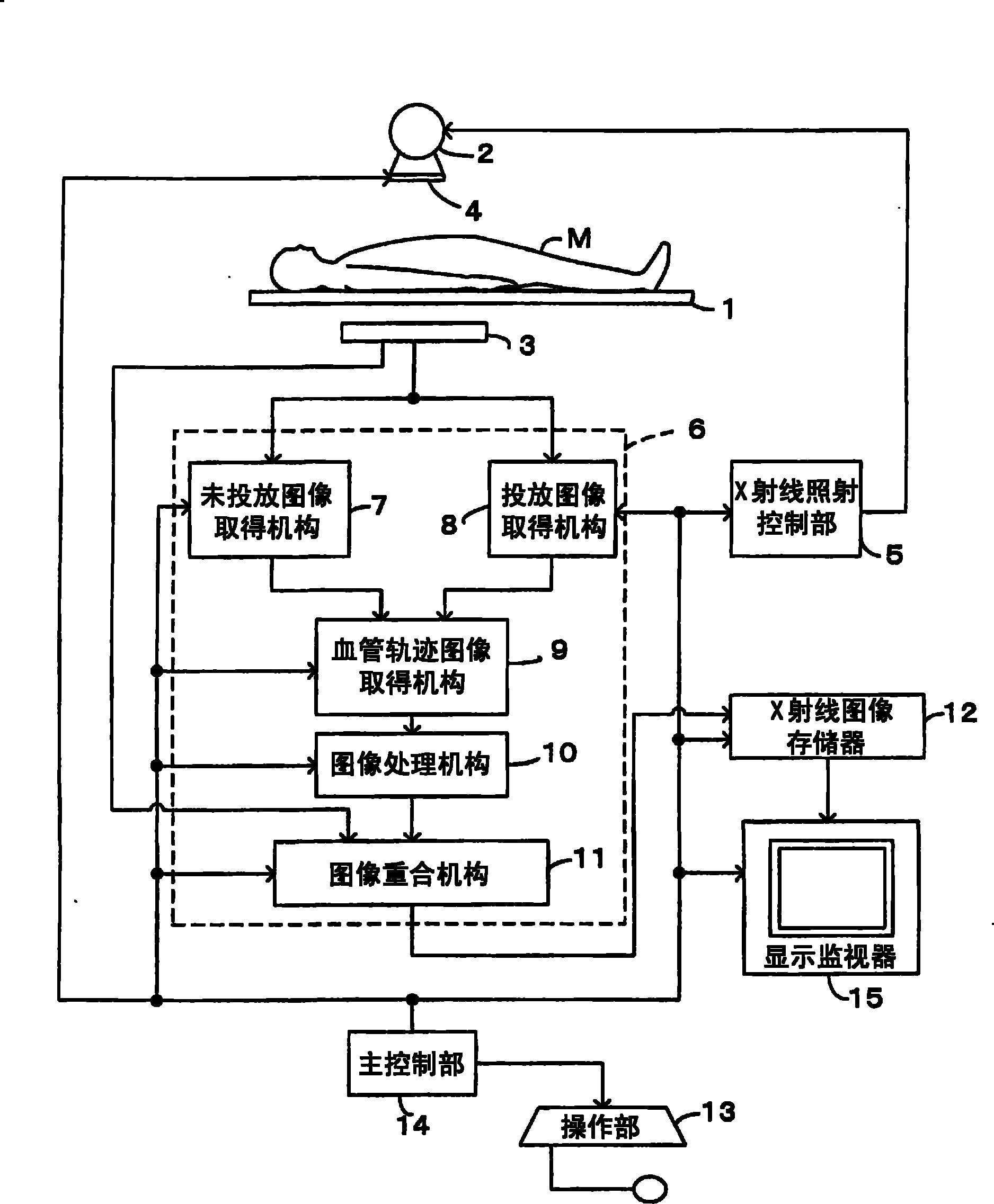
  • Figure 1
    Figure 1
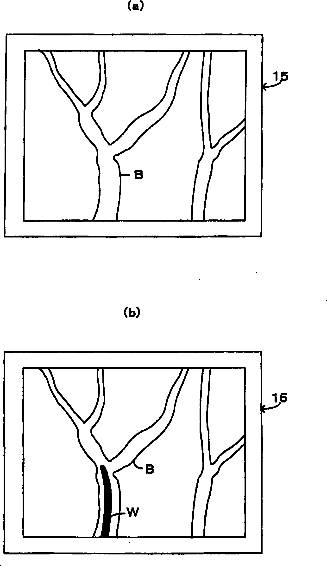
  • Figure 2
    Figure 2
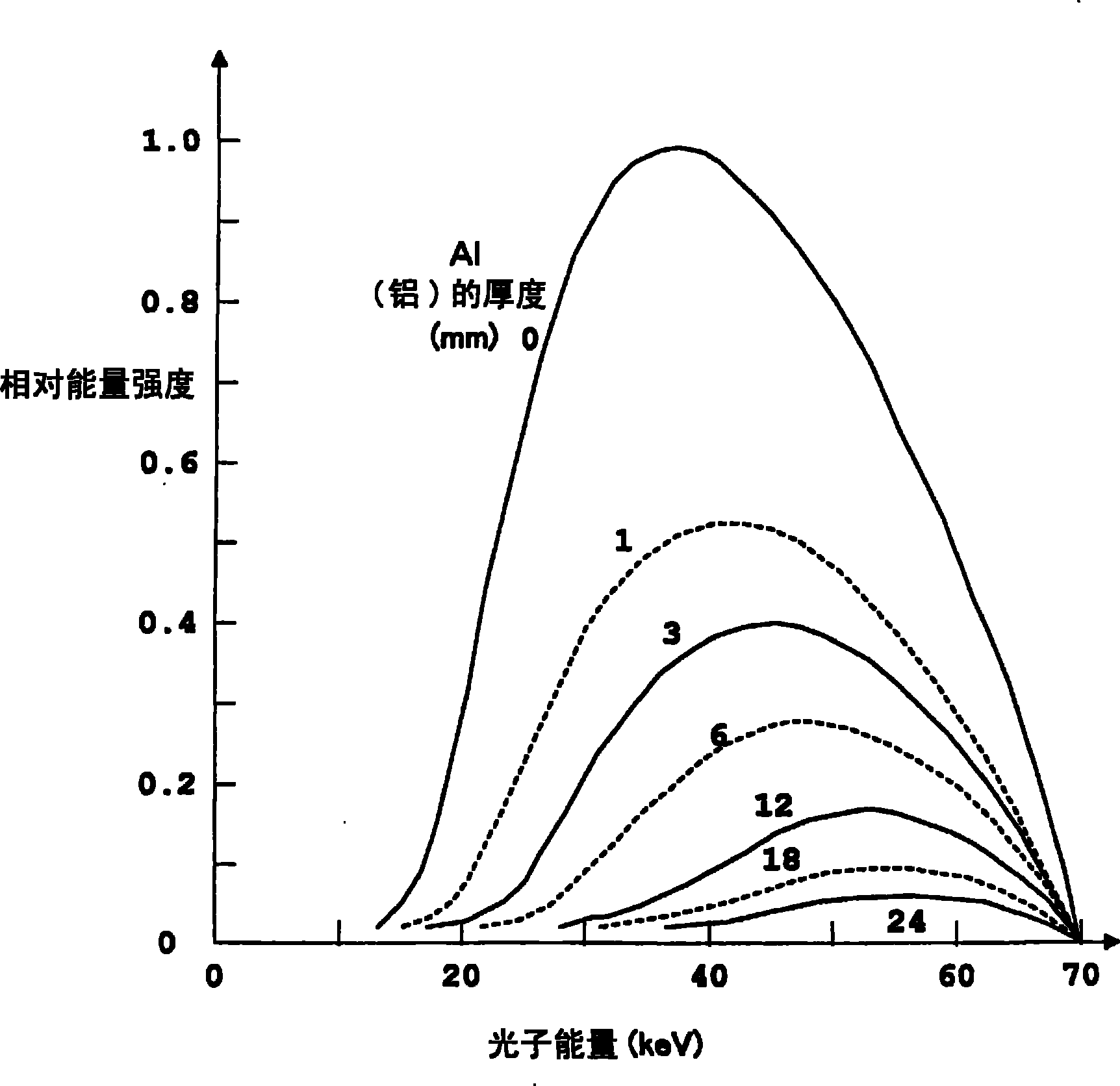
  • Figure 3
    Figure 3
Patent Text Reader

Abstract

X-rays are applied without administrating an angiographic contrast agent, unadministration image acquiring means (7) acquires an unadministration image of an angiographic contrast agent unadministrated state, X-rays are applied after administrating an angiographic contrast agent, and an administration image acquiring means (8) acquires an administration image of an angiographic contrast agent administered state. The acquired unadministration and administration images are superimposed on one the other by blood vessel trace image acquiring means (9), the image data on the DSA images obtained inthe angiographic contrast agent unadministrated and administrated states is processed, and thus a blood vessel trace image is acquired. The acquired blood vessel trace image is black-white inverted by image processing means (10), and blood vessels are displayed in white. A beam hardening filter (4) is inserted. While inserting a guide wire (W) in the blood vessel, X-ray fluoroscopic imaging is performed. The X-ray fluoroscopic image and the blood vessel trace image are superimposed on one the other by image superimposing means (11), and the acquired superimposed image is displayed on a display monitor (15).
Need to check novelty before this filing date? Find Prior Art

Description

technical field

[0001] The present invention relates to an X-ray diagnostic apparatus including: an X-ray irradiation mechanism such as an X-ray tube that irradiates an object with X-rays; Signal X-ray detectors and other X-ray detection mechanisms. In particular, it relates to a technique for generating road map images. Background technique

[0002] An example of using an X-ray fluoroscopy device as an X-ray diagnostic device will be described. Conventionally, the following configurations have been known as techniques for generating road map images of such X-ray fluoroscopy devices.

[0003] That is, with X-ray irradiation from the X-ray tube to the subject, an X-ray detection signal is output from the flat-panel X-ray detector, and the transmitted X-ray of the subject is obtained by the data processing unit based on the X-ray detection signal. The corresponding X-ray image is stored in the X-ray image memory, and the X-ray image and the operation menu necessary for X-ra...

Examples

Embodiment Construction

[0040] Hereinafter, embodiments of the present invention will be described in detail with reference to the drawings.

[0041] figure 1 It is an overall schematic configuration diagram showing an X-ray diagnostic apparatus according to an embodiment of the present invention. An X-ray tube as an X-ray irradiation mechanism for irradiating X-rays is installed above the table 1 across the table 1 on which the subject M is mounted. 2. On the other hand, an X-ray detector 3 as an X-ray detection mechanism for detecting X-rays irradiated from the X-ray tube 2 and transmitted through the subject M is provided under the table top 1 . In the present embodiment, an X-ray fluoroscopy device is used as an example of an X-ray diagnostic device for description.

[0042] In front of the X-ray irradiation direction of the X-ray tube 2, a beam quality control mechanism that can switch between the inactive state and the active state to control the energy distribution of the X-rays irradiated fr...