Method for quantitative digital microscopic phase contrast imaging

A phase-contrast imaging and digital microscopy technology, applied in microscopes, optics, instruments, etc., can solve problems such as complex structures and poor noise robustness, and achieve the effects of rapid quantitative phase analysis and rapid quantitative microscopic phase contrast imaging

Inactive Publication Date: 2009-11-11
BEIHANG UNIV
View PDF0 Cites 12 Cited by
  • Summary
  • Abstract
  • Description
  • Claims
  • Application Information

AI Technical Summary

Problems solved by technology

However, the system still needs a complex and expensive phase difference tube to acquire the microscopic image sequence; in addition, the system needs to make str

Method used


Image

  • Method for quantitative digital microscopic phase contrast imaging
  • Method for quantitative digital microscopic phase contrast imaging
  • Method for quantitative digital microscopic phase contrast imaging

Examples


Embodiment Construction

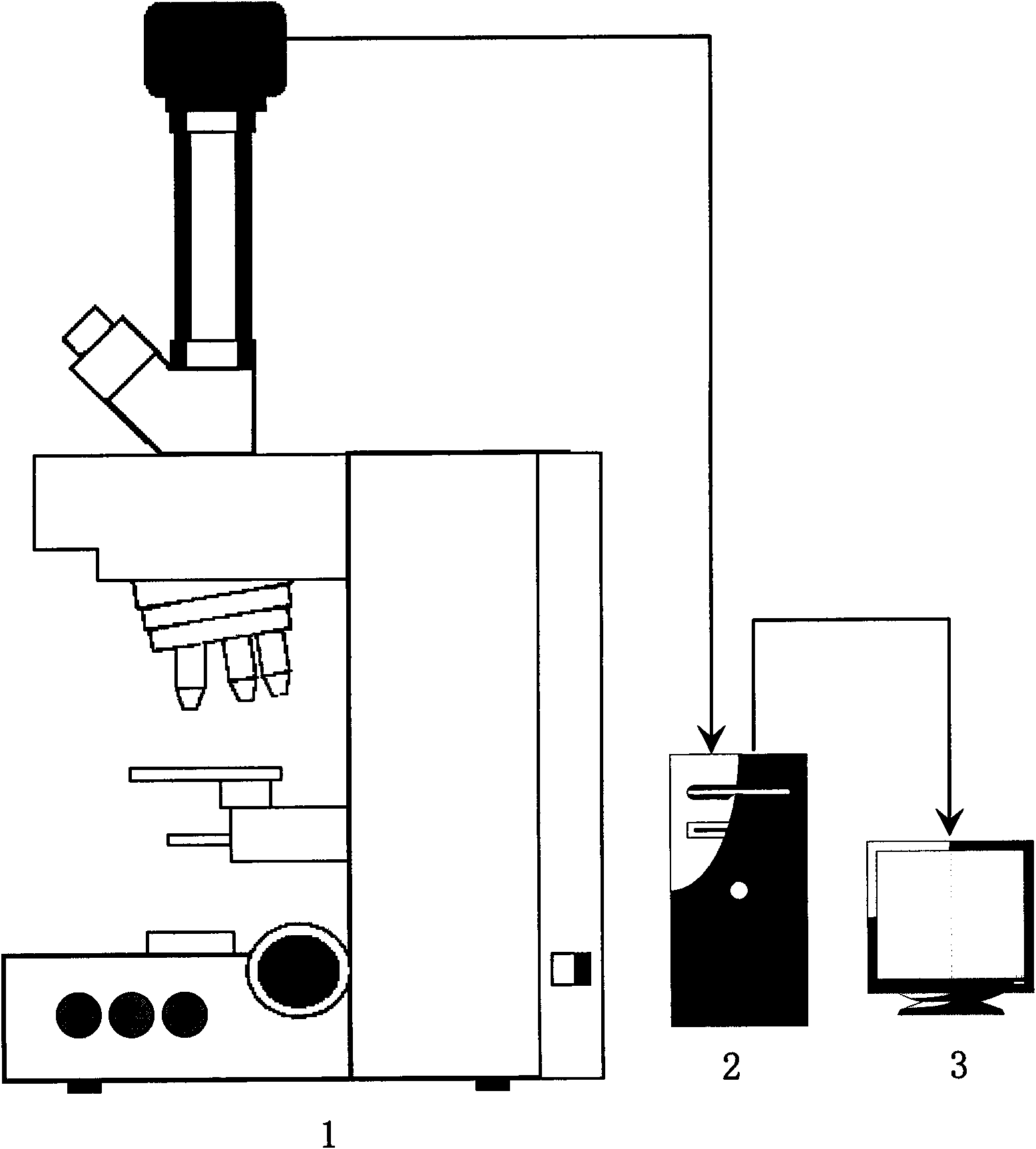
[0063] The present invention implements required equipment condition to be: 1. a common bright-field optical transmission digital microscope system; 2. digital phase recovery computer is common PIV3.4Ghz 1G RAM microcomputer, as attached figure 1 ③The physical parameters of the digital microscope are: objective lens 40X, camera model M1500, camera CCD chip pixel size 4.65 microns, trinocular camera connector 0.65X, image resolution 696X520, and scale bar 0.35 microns / pixel.

[0064] The environmental conditions required for the specific implementation of the present invention are: an ordinary bright field optical transmission digital microscope imaging environment.

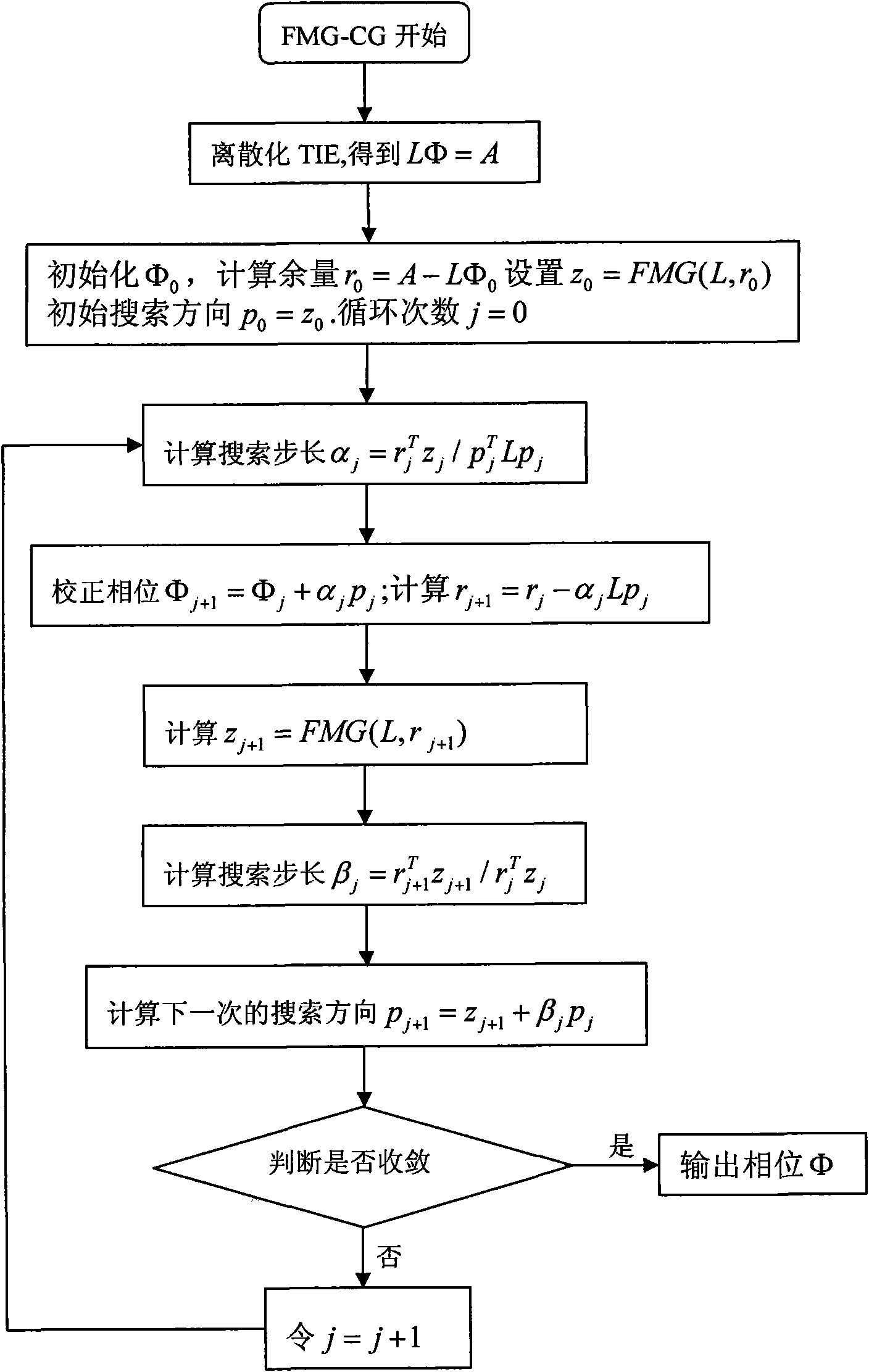
[0065] The present invention is a method for quantitative digital microscopic phase contrast imaging, the process of quantitative phase recovery in the method is as attached figure 2 shown.

[0066] The specific implementation steps of the method are as follows:

[0067] Step 1: Establish the light intensity pr...


PUM


Abstract

The invention provides a method for quantitative digital microscopic phase contrast imaging, which is realized on a normal digital optical microscope platform and a general personal computer and comprises the following four steps of: step one: establishing a light intensity transmission equation and carrying out discretization treatment on the equation; step two: designing a discretized light intensity transmission equation numerical algorithm; step three: collecting the light intensity data; and step four: carrying out quantitative phase recovery and phase contrast imaging. The method is on the normal digital optical microscope, needs not to increase complex optical parts, realizes the phase contrast imaging on the basis of phase recovery numerical algorithm, improves the contrast of the transparent or semitransparent sample imaging, and quickly realizes quantitative microscopic phase contrast imaging and quantitative phase analysis. The method is also applicable to a microscopic imaging system of other modes and has wide practical values and application prospects in the technical field of microscopic optical imaging.

Description

(1) Technical field [0001] The invention relates to a method for quantitative digital microscopic phase-contrast imaging. Specifically, it is a method for realizing phase-contrast imaging based on numerical algorithms on an ordinary digital optical microscope platform without adding complicated optical components, and improving transparency or translucency. The invention relates to contrast of transparent sample imaging, which belongs to the technical field of microscopic optical imaging. (2) Background technology [0002] In biological and medical research, it is often necessary to observe some very important transparent organisms or tissues (such as cells). Since the intensity of light does not change significantly after passing through such objects, but the phase will shift, when imaging with an ordinary transmission microscope , the structure of the organism will disappear into the medium around it. In order to observe and study such organisms under a light microscope, ...

Claims


Application Information

Patent Timeline
IPC IPC(8): G02B21/00
Inventor 薛斌党郑世玲
Owner BEIHANG UNIV
Why Eureka
Try Eureka