Hepatic segment volume measuring method based on CT reinforcing scan technique

A liver segmentation and enhanced scanning technology, applied in the field of medical image processing, can solve the problems of poor correlation of liver anatomical segmentation, segmentation errors, shape, size, number and anatomical variation

Inactive Publication Date: 2009-12-16
厦门强本科技有限公司 +1
View PDF0 Cites 36 Cited by
  • Summary
  • Abstract
  • Description
  • Claims
  • Application Information

AI Technical Summary

Problems solved by technology

However, because the branches of the portal vein have great anatomical variations in shape, size, number, etc.; at the same time, the boundary between segments is not a simple plane but irregular waves, so the traditional CT section segmentation method does not fully match the actual anatomy.
That is, according to traditional radiological standards, the correlation between CT slice liver segmentation and real liver anatomical segmentation is poor. According to research, the segmentation error of axial CT slice reaches 40%.
Therefore, the traditional CT slice segmentation method must be modified

Method used


Image

  • Hepatic segment volume measuring method based on CT reinforcing scan technique
  • Hepatic segment volume measuring method based on CT reinforcing scan technique
  • Hepatic segment volume measuring method based on CT reinforcing scan technique

Examples


Embodiment Construction

[0032] The following embodiments will further illustrate the present invention in conjunction with the accompanying drawings.

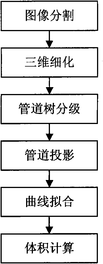
[0033] figure 1 Provide the flow chart of the embodiment of the present invention, its specific steps are as follows:

[0034] Step 1. Image segmentation:

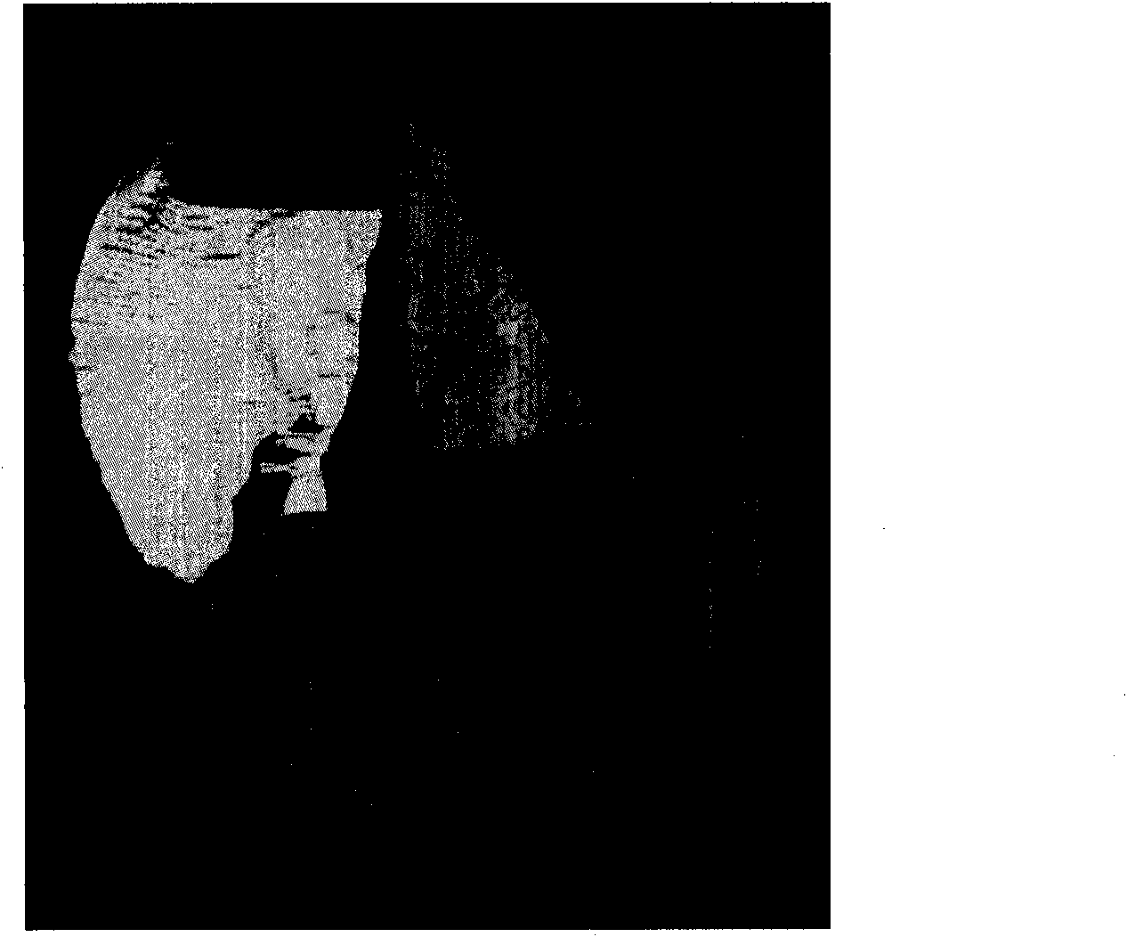
[0035] The purpose of image segmentation is to process the data obtained after CT scanning to obtain the surface contour of the liver organ and the pipeline area inside the liver.

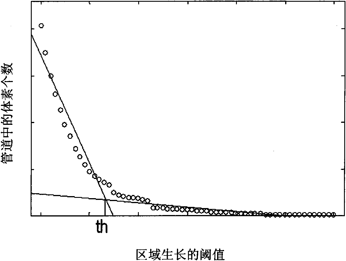
[0036] At present, there are many algorithms for image segmentation, but none of them can be fully applied to segment the surface contour of the liver. For the sake of reliability, to improve the accuracy of image segmentation, the surface contour of the liver is segmented by manual interaction. The contrast agent is injected before the CT examination. During CT imaging, the CT value of the intrahepatic duct is much higher than the CT value of the liver parenchyma. The threshold segmentation technology can be used to...


PUM


Abstract

The invention provides a hepatic segment volume measuring method based on a CT reinforcement scan technique, and relates to a method for processing medical images. The method comprises the following steps of: adopting an image processing algorithm to process data acquired after CT scan, and acquiring the surface profile of a liver organ and an intrahepatic vessel area; acquiring the central line of the hepatic vessel; processing the central line of the hepatic vessel into a tree structure, namely the number of nodes is equal to the number of edges plus one; projecting the liver data on a projection plane, and converting a three-dimensional liver into a two-dimensional classification in segments; processing the central lines of the blood vessels into smooth central curves of the blood vessels; and dividing the liver according to the central curves of the blood vessels, and working out the volume of each hepatic segment. The liver data acquired based on the CT reinforcement scan technique is subjected to Couinaud hepatic segment division, so that volumes of hepatic segments are acquired, and the method has great clinical significance.

Description

technical field [0001] The invention relates to a medical image processing method, in particular to a liver segmental volume measurement method based on CT enhanced scanning technology. Background technique [0002] CT (Computerized Tomography) is widely used in the field of medical clinical diagnosis. Enhanced CT scan is to scan the image after injecting contrast agent into the patient, so as to obtain images of the hepatic artery, hepatic vein, and portal vein in the liver. [0003] At present, with the rapid development of liver surgery, hepatic segment, sub-segment and even wedge resection have become possible. Therefore, there is an urgent need for accurate preoperative localization and liver segmentation of liver lesions. At present, the Couinaud liver segment division method (Soyer P. Segmental anatomy of the liver utility of anomenclature accepted worldwide [J]. AJR, 1993, 161(3): 572-573) is widely used in the world, which is based on the portal vein sheath system ...

Claims


Application Information

Patent Timeline
Patent Type & Authority Applications(China)
IPC IPC(8): A61B5/107G06T5/00
Inventor 王博亮黄晓阳黄绍辉
Owner 厦门强本科技有限公司
Why Eureka
Try Eureka