Method for analyzing pore structure of solid material based on microscopic image

A solid material and pore structure technology, applied in 3D image processing, image data processing, permeability/surface area analysis, etc.

Inactive Publication Date: 2010-02-03
TAIYUAN UNIV OF TECH
View PDF0 Cites 69 Cited by
  • Summary
  • Abstract
  • Description
  • Claims
  • Application Information

AI Technical Summary

Problems solved by technology

[0005] The present invention analyzes the pore structure of solid materials based on microscopic images, and aims to overcome the deficiencies and defects of the prior art and solve unsolvable problems in the prior art. According to CT, it has the ability to detect the internal structure of solid materials non-destructively and generate

Method used


Image

  • Method for analyzing pore structure of solid material based on microscopic image
  • Method for analyzing pore structure of solid material based on microscopic image
  • Method for analyzing pore structure of solid material based on microscopic image

Examples


Embodiment approach 1

[0032] The mudstone is scanned by the micro-CT X-ray imaging system, and the computer system reconstructs the scanned CT image to generate a single CT cross-sectional image. Digital image processing and three-dimensional reconstruction are performed based on the micro-CT image. The purpose is to calculate Mudstone pore size, porosity and their variation. The calculation process is:

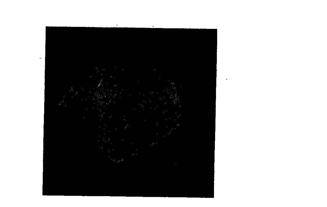
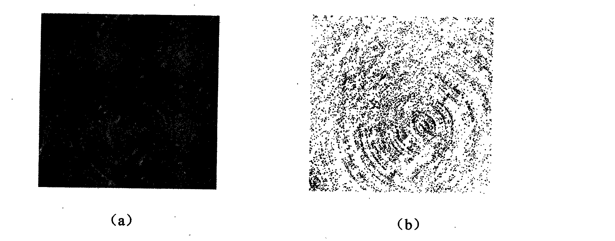
[0033] I. Acquisition of a single cross-sectional image of micro-CT. The micro-CT single image comes from the micro-CT test system, figure 1 It is a randomly selected cross-sectional image in bmp bitmap format, the image type is index image, the image size is 2001×2001 pixels, the number of bits per pixel is 8 bits, and the grayscale range is 0 to 255. The size is 1.26 μm, that is, the pore diameter is 1.26 μm. The black dots in the figure indicate the lower density of the material, the white dots indicate the higher density of the material, and the change from black to white indicates the dens...

Embodiment approach 2

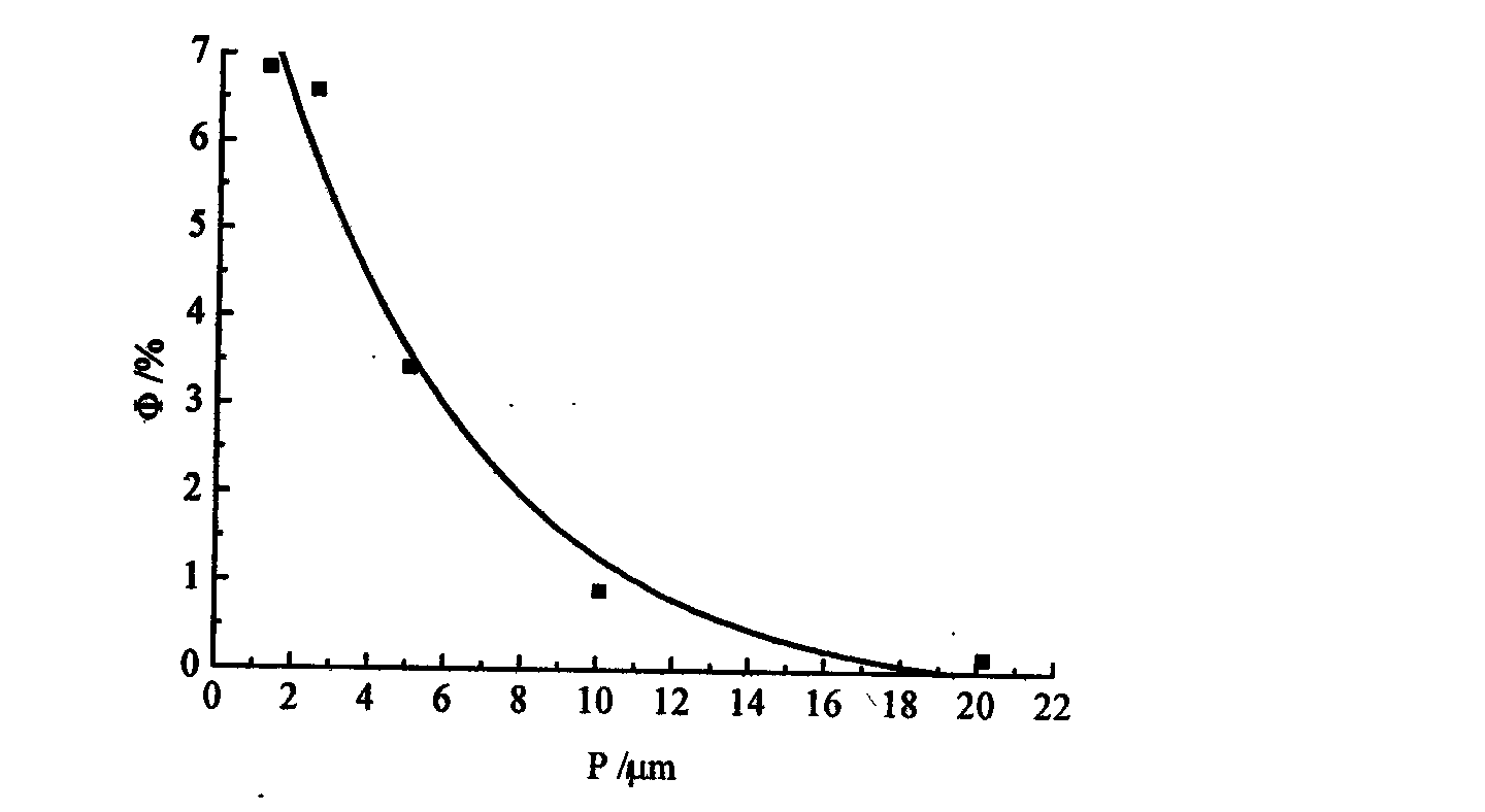
[0044] Embodiment 2: The solid material coal is scanned by the micro-CT X-ray imaging system, and the pore structure of the coal is analyzed. The pore size is in the range of 0.8 μm to 7 μm, and the porosity is in the range of 22% to 4%. The law of decrement. Others are the same as Embodiment 1.

Embodiment approach 3

[0045] Embodiment 3: The solid material sandstone is scanned by the micro-CT X-ray imaging system, and the pore structure of the sandstone is analyzed. The pore size is in the range of 0.4 μm to 4 μm, and the porosity is in the range of 2% to 14%. The law of decrement. Others are the same as Embodiment 1.


PUM


Abstract

The invention provides a method for analyzing the pore structure of solid material based on microscopic image, belonging to the technical field for analyzing the pore structure of the solid material.The method is characterized of: obtaining the CT single cross section image of the solid material by microscopic CT scanning; using computer language to digitally image process the CT single cross section image; taking the pixel side of the image as the size of hole diameter; computing the hole diameter of the solid material, porosity and change regularity of the hole diameter and the porosity based on the microscopic CT single image; selecting the CT single image after processing a plurality of digital image; generating a CT image sequence; three dimensionally rebuilding the CT single image with a volume rendering algorithm in a visual rebuilding algorithm; generating the three dimensional digital image of the solid material; and computing the hole diameter of the solid material, the porosity and the change regularity of the hole diameter and the porosity based on the microscopic CT single image. The method is widely used for analyzing and computing the hole size and the porosity of the solid material under the various hole sizes of the solid material.

Description

technical field [0001] The invention discloses a method for analyzing the pore structure of a solid material based on a microscopic image, belonging to the technical category of analyzing the pore structure of a solid material, and specifically relates to a method for analyzing the pore structure of a solid material based on a micro-CT image. Background technique [0002] In the prior art for analyzing the pore structure of solid materials, there are mainly three kinds of low-temperature nitrogen adsorption method, mercury intrusion method and traditional density method. The pore diameters of solid materials measured by the existing technology have a limited range, and the distribution range of the pore diameters is from nanometer to millimeter, a total of 6 orders of magnitude. The existing measuring instruments and methods cannot measure all the pore structures. Both have been measured, so the porosity of solid materials measured by different methods is different, and its ...

Claims


Application Information

Patent Timeline
IPC IPC(8): G01N15/08G06T15/00G06T17/00
Inventor 王东康天合柴肇云李义宝杨永康
Owner TAIYUAN UNIV OF TECH
Why Eureka
Try Eureka