Automatic segmentation method for retinal nerve fiber layer in OCT image of ocular fundus

An optic nerve fiber layer and automatic segmentation technology, applied in the field of image processing, can solve the problems of reducing the inherent feature description ability of biological tissue, and can not solve the problem of data noise, and achieve the effect of accurate segmentation and extraction, and rich information.

Inactive Publication Date: 2010-03-31
SHENZHEN GRADUATE SCHOOL TSINGHUA UNIV
View PDF0 Cites 16 Cited by
  • Summary
  • Abstract
  • Description
  • Claims
  • Application Information

AI Technical Summary

Problems solved by technology

Using this simplified grayscale image will reduce the ability to describe the inherent characteristics of biological tissues during processing and analysis, and at the same time cannot solve the problem of data noise well

Method used


Image

  • Automatic segmentation method for retinal nerve fiber layer in OCT image of ocular fundus
  • Automatic segmentation method for retinal nerve fiber layer in OCT image of ocular fundus
  • Automatic segmentation method for retinal nerve fiber layer in OCT image of ocular fundus

Examples


Embodiment Construction

[0033] The features and advantages of the present invention will be described in detail with reference to the accompanying drawings.

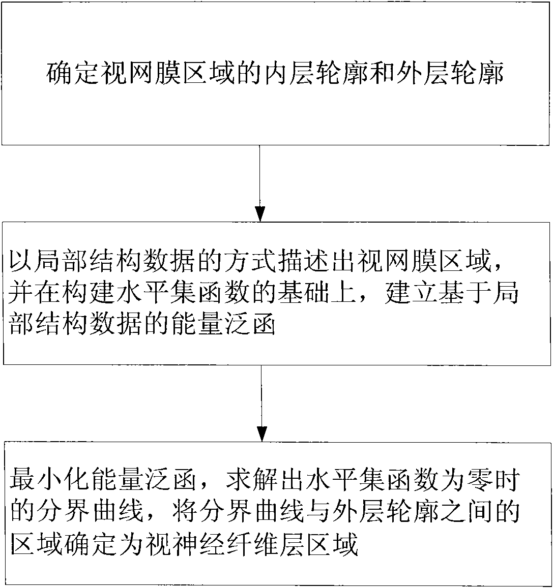
[0034] Please refer to figure 2 , the flow of an embodiment of the method for automatic segmentation of the optic nerve fiber layer is as follows.

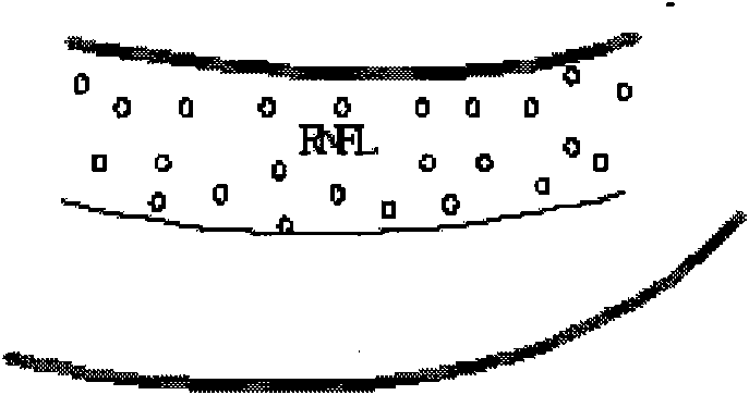
[0035] Assuming that the contours of the inner and outer layers of the retinal region are already known, they are expressed as: u(s)=[x(s), y(s)], v(s)=[x(s), y(s)], the boundary of RNFL The curve is m(s)=[x(s), y(s)], the area between u and v is represented by Ω, and the area of ​​RNFL is represented by Ω 1 Indicates that the non-RNFL area is represented by Ω 2 express. Use p(x, y) to represent the local structure data of the OCT image.

[0036] According to the level set theory, the energy norm based on non-Euclidean data p(x, y) is established:

[0037]

[0038] Among them, d( ) is the distance function, which describes the difference between different local structure data, for Ω 1 m...


PUM


Abstract

The present invention discloses an automatic segmentation method for retinal nerve fiber layer in OCT image of ocular fundus, comprising the following steps: A. determining the inner layer outline u(s) of the retinal region as u(s)=[x(s), y (s)], and the outer layer outline v(s) as v(s)=[x(s), y(s)]; B. describing the retinal region in local structure data p (x, y) mode, and constructing the energy function (the formula is shown above) based on the local structure data p (x, y) after constructing the level set function phi; and C. minimizing the energy function, calculating the boundary curvem as m= {(x, y) / phi (x, y) = 0} while phi (x, y) = 0, and defining the region between the boundary curve m(s) and the outer layer outline v(s) as the retinal nerve fiber layer region. According to the invention, retinal nerve fiber layer in OCT image of ocular fundus can be segmented reliably and accurately.

Description

【Technical field】 [0001] The invention relates to image processing, in particular to a method for automatically segmenting optic nerve fiber layers of fundus OCT images. 【Background technique】 [0002] Optical coherence tomography (Optical Coherence Tomography, OCT) is an imaging technology that has developed rapidly in recent years. High rate, high sensitivity, non-invasive, non-contact and other characteristics, it occupies a place in many imaging methods, and has a good development prospect, and has attracted more and more attention. OCT uses the weak coherence characteristics of broadband light sources to perform high-resolution tomographic imaging of the internal microstructure of biological tissues by measuring the interference signal of backscattered light from the sample. Compared with traditional optical microscopy, OCT utilizes the weak coherence of broadband light to suppress background energy, and can obtain a resolution (μm level) that is 1 to 2 orders of magni...

Claims


Application Information

Patent Timeline
Patent Type & Authority Applications(China)
IPC IPC(8): G06T5/00A61B3/12
Inventor 卢宗庆杨帆廖庆敏
Owner SHENZHEN GRADUATE SCHOOL TSINGHUA UNIV
Why Eureka
Try Eureka