Reservoir eluting stent

An internal stent and implantable technology, applied in the direction of stents, dilators, and medical devices, can solve problems such as vascular obstruction and embolism

Active Publication Date: 2015-02-11
CARDINAL HEALTH SWITZERLAND 515 GMBH
View PDF23 Cites 0 Cited by
  • Summary
  • Abstract
  • Description
  • Claims
  • Application Information

AI Technical Summary

Problems solved by technology

Such detachment may again lead to adverse outcomes including embolization of coating fragments (leading to vascular occlusion)

Method used


Image

  • Reservoir eluting stent
  • Reservoir eluting stent
  • Reservoir eluting stent

Examples


example 1

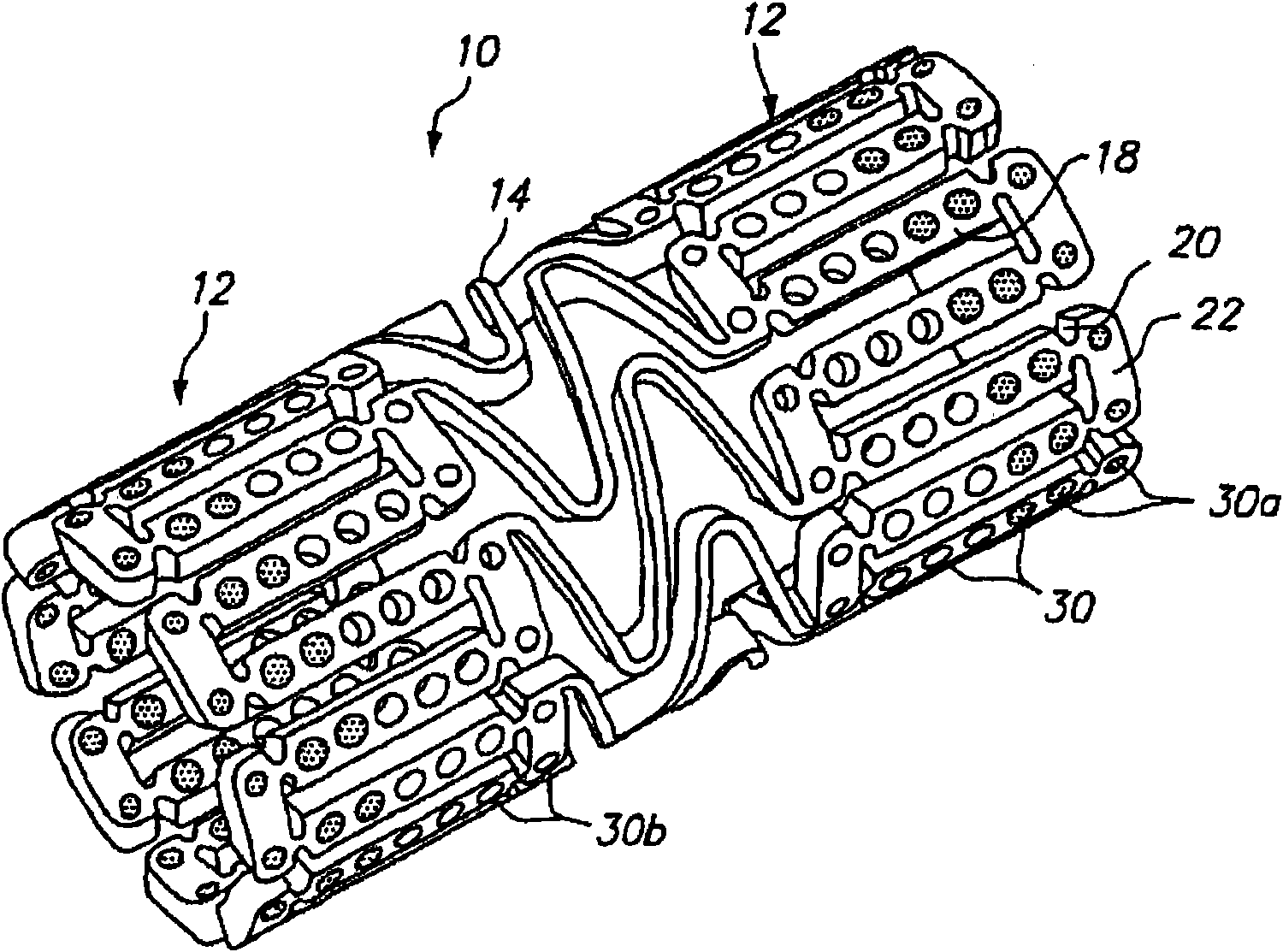
[0075] Figure 7 A dual drug stent 700 is illustrated with an anti-inflammatory agent and an anti-proliferative agent delivered from different orifices of the stent so that the two drugs have independent release kinetics specifically designed to match the biological process of restenosis. According to this example, the dual drug stent comprises a combination of the anti-inflammatory agent pimecrolimus in the first set of openings 710 and the anti-proliferative agent paclitaxel in the second set of openings 720 . Each agent is provided in the matrix material within the pores of the stent in a specific embedded arrangement designed to achieve Figure 8 Release kinetics shown. Each drug is delivered primarily along the lumen wall to treat restenosis.

[0076] Such as Figure 7As shown, pimecrolimus is positioned in the stent for targeted delivery to the luminal side of the stent by using a barrier layer 712 on the luminal side of the pores. The barrier layer 712 is formed of ...

example 2

[0082] According to this example, the dual drug stent contains Gleevec in a first set of openings 710 and the antiproliferative agent paclitaxel in a second set of openings 720 . Each agent is provided in the matrix material within the pores of the stent in a specific embedded arrangement designed to achieve Figure 8 Release kinetics shown.

[0083] Gleevec is delivered with a two-phase release, including an initial high-speed release on the first day followed by a slow release for 1 to 2 weeks. The first phase of Gleevec release is to deliver approximately 50% of the drug load within approximately the first 24 hours. The second phase of release is to deliver the remaining 50% in about 1-2 weeks. Such as Figure 8 As shown in and described in Example 1 above, paclitaxel was loaded into opening 720 in such a way that the resulting release kinetics were substantially linear after about an initial 24 hours.

[0084] Depending on the size of the stent, the amount of drug deli...

example 3

[0086] According to this example, the dual drug stent comprises a combination of PKC-412 (a cell growth regulator) in a first set of openings and the antiproliferative agent paclitaxel in a second set of openings. Each agent is provided in the matrix material within the pores of the stent in a specific embedded arrangement designed to achieve release kinetics as described below.

[0087] PKC-412 is delivered at a substantially constant release rate after about the first 24 hours, over a period of about 4 weeks to 16 weeks, preferably 6 weeks to 12 weeks. Paclitaxel is loaded into the openings in such a way that the resulting release kinetics are substantially linear after about an initial 24 hours, with a release period of about 4 to 16 weeks, preferably about 6 to 12 weeks.

[0088] Depending on the size of the stent, the amount of drug delivered will vary. For a 3 mm x 6 mm scaffold, the amount of PKC-412 is about 100 micrograms to about 400 micrograms, preferably about 150...


PUM

PropertyMeasurementUnit
diameteraaaaaaaaaa

Abstract

An expandable medical device includes a plurality of elongated struts, forming a substantially cylindrical device which is expandable from a first diameter to a second diameter. A plurality of different beneficial agents may be loaded into different openings within the struts for delivery to the tissue. For treatment of conditions such as restenosis, different agents are loaded into different openings in the device to address different biological processes involved in restenosis and are delivered at different release kinetics matched to the biological process treated. The different agents may also be used to address different diseases from the same drug delivery device. In addition, anti-thrombotic agents may be affixed to at least a portion of the surfaces of the medical device for the prevention of sub-acute thrombosis. Primer layers may be used to ensure that the different agents remain affixed to the device as well as to each other.

Description

technical field [0001] This application claims priority to US Provisional Application No. 61 / 148,610, filed January 30, 2009. [0002] The present invention relates to tissue supporting medical devices, and more particularly to non-removable expandable devices that are implanted in a body cavity of a living animal or human to support and maintain organs The beneficial agent is delivered to the opening of the intervention site as well as the antithrombotic agent surface coating. Background technique [0003] In the past, permanent or biodegradable devices have been developed for implantation in body passages to keep the passage open. These devices are usually introduced percutaneously and delivered transluminally until positioned at the desired location. The device is then expanded mechanically (eg, by expanding a mandrel or balloon disposed within the device), or self-expanding by releasing stored energy upon physical activation. Once expanded within a body cavity, the de...

Claims


Application Information

Patent Timeline
Patent Type & Authority Patents(China)
IPC IPC(8): A61F2/82A61M31/00A61M29/00A61F2/91
CPCA61L2300/416A61L31/16A61L31/10A61F2250/0068A61F2/91A61L2300/604A61P7/02A61P9/10A61P37/06C08L67/04
Inventor S·普赖斯J·R·L·斯坦利A·卢克Y·-P·孙J·杜利L·-G·盖R·科瓦尔西克D·博德里G·斯蒂斯-布拉利C·罗杰斯
Owner CARDINAL HEALTH SWITZERLAND 515 GMBH
Why Eureka
Try Eureka