
Cardiovascular x-ray diagnostic system

An X-ray and X-ray tube technology, applied in the field of X-ray diagnostic system for circulators, can solve problems such as inability to collect photographic images

Inactive Publication Date: 2010-09-22
KK TOSHIBA +1
View PDF6 Cites 3 Cited by
  • Summary
  • Abstract
  • Description
  • Claims
  • Application Information

AI Technical Summary

Problems solved by technology

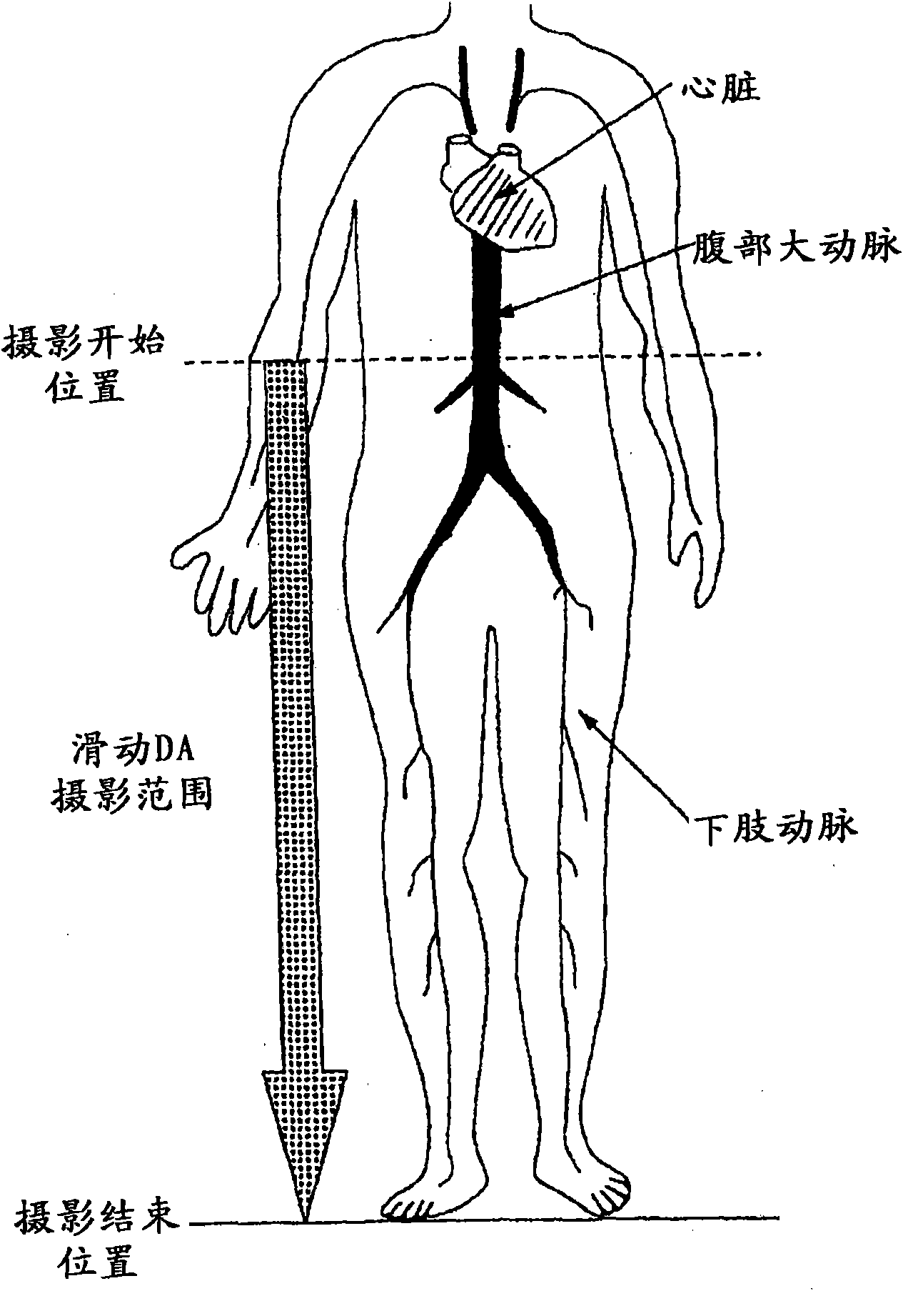
However, the operator sometimes forgets to switch the imaging program or imaging condition parameters before performing sliding DA imaging of the lower limbs, and in this case, there is a problem that appropriate imaging images may not be collected.

Method used


Image

  • Cardiovascular x-ray diagnostic system
  • Cardiovascular x-ray diagnostic system
  • Cardiovascular x-ray diagnostic system

Examples


Embodiment Construction

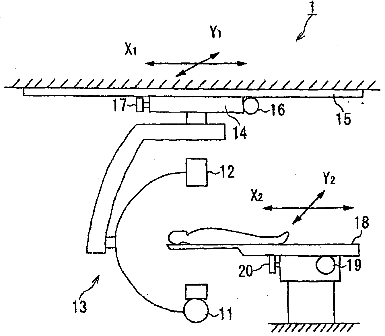
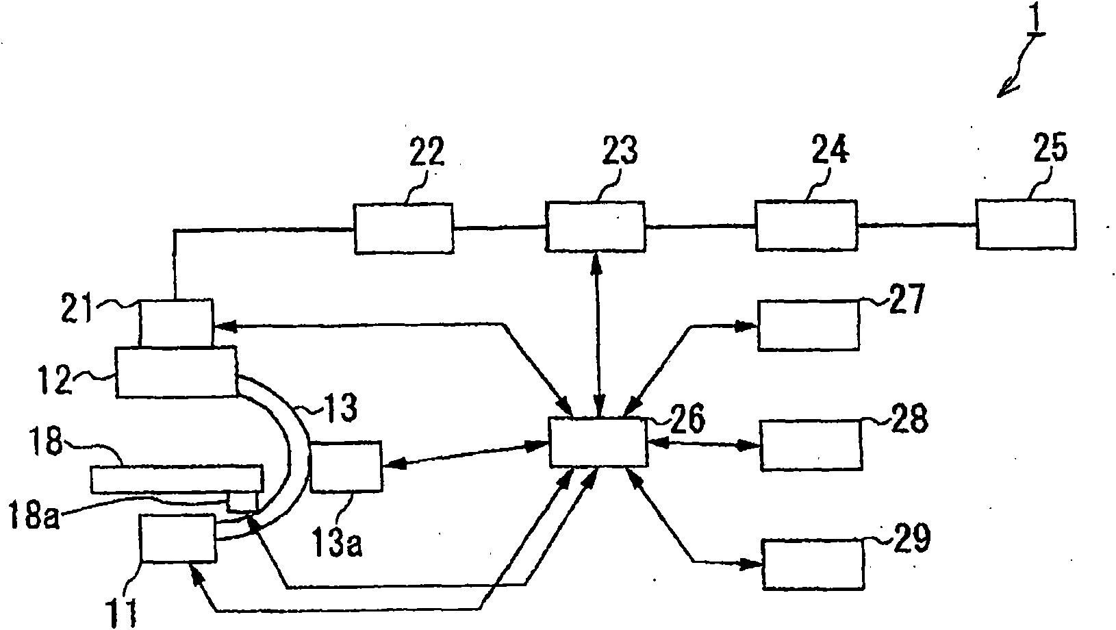
[0020] Embodiments of the X-ray diagnostic system for a circulator according to the present invention will be described in detail with reference to the drawings. figure 1 It is a figure roughly showing the whole structure of the X-ray diagnostic system 1 for circulators of this invention, figure 2 It is a configuration diagram showing the X-ray diagnostic system 1 for a circulator of the present invention.

[0021] X-ray diagnostic system for circulator 1 such as figure 1 and figure 2 As shown, it is equipped with: an X-ray generating device 11 that generates and irradiates X-rays; an X-ray imaging device 12 that detects X-rays transmitted through the subject and converts them into electrical signals for imaging; and The arm 13 of the X-ray generating device 11 and the X-ray imaging device 12 is held securely.

[0022] The X-ray generator 11 includes an X-ray tube, a limiter that limits the irradiation range of X-rays, a high-voltage device that applies a high voltage to ...


PUM


Abstract

A cardiovascular x-ray diagnostic system capable of taking radiographs at different positions by moving at least one of a tabletop on which a subject is placed and a radiographic system equipped with an x-ray tube and an x-ray detector includes: an acquisition unit configured to acquire a radiography position from a position of the tabletop and a position of the radiographic system; a reference position identification unit configured to identify a reference radiography position; and a radiographic unit configured to take radiographs by changing a radiographic parameter based on a current radiography position acquired by the acquisition unit and the reference radiography position.

Description

technical field [0001] The present invention relates to an X-ray diagnostic system for a circulator that sequentially selects and sets an optimal imaging program or imaging condition parameter according to an imaging site when imaging is performed while moving a target site. Background technique [0002] In recent years, X-ray diagnostic systems for circulators have been used as devices that support not only routine examinations for existing diagnoses but also treatment (IVR: Interventional Radiology) using catheters in real time. In catheter-based treatment (IVR) and examination for coronary artery stenosis, angiographic confirmation of lower extremity arteries is sometimes performed after a series of coronary angiography and treatment due to the risk of concurrent lower extremity arterial stenosis. In this imaging, since it is desired to perform imaging over a wide range as much as possible by injecting a contrast agent in order to reduce the amount of contrast agent, a me...

Claims


Application Information

Patent Timeline
Patent Type & Authority Applications(China)
IPC IPC(8): A61B6/00
CPCA61B6/544A61B6/547A61B6/4441A61B6/542A61B6/503A61B6/504A61B6/545A61B6/04A61B6/4464
Inventor 小泽政广石川贵之渡部勇一郎
Owner KK TOSHIBA
Learn More