
Positron emission tomography apparatus and nuclear medical image generating method

A positron, incident direction technology, used in measuring devices, scientific instruments, computer tomography scanners, etc., can solve problems such as cost

Inactive Publication Date: 2010-10-20
KK TOSHIBA +1
View PDF4 Cites 11 Cited by
  • Summary
  • Abstract
  • Description
  • Claims
  • Application Information

AI Technical Summary

Problems solved by technology

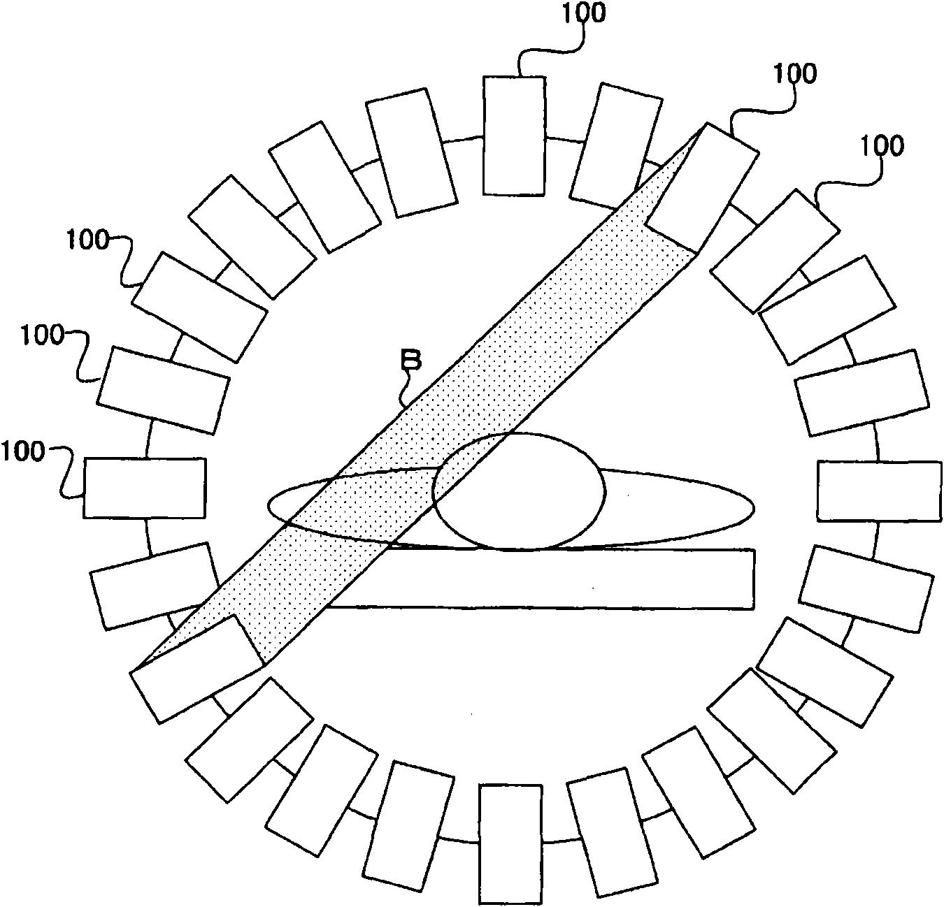
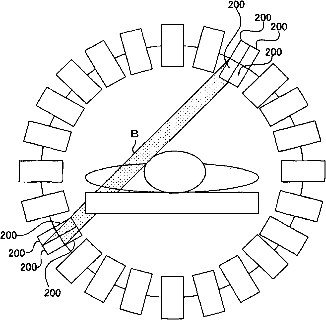
[0009] However, since the DOI detector is equipped with a plurality of scintillators 200 in the depth direction, it costs a lot of money.

Method used


Image

  • Positron emission tomography apparatus and nuclear medical image generating method
  • Positron emission tomography apparatus and nuclear medical image generating method
  • Positron emission tomography apparatus and nuclear medical image generating method

Examples


Embodiment Construction

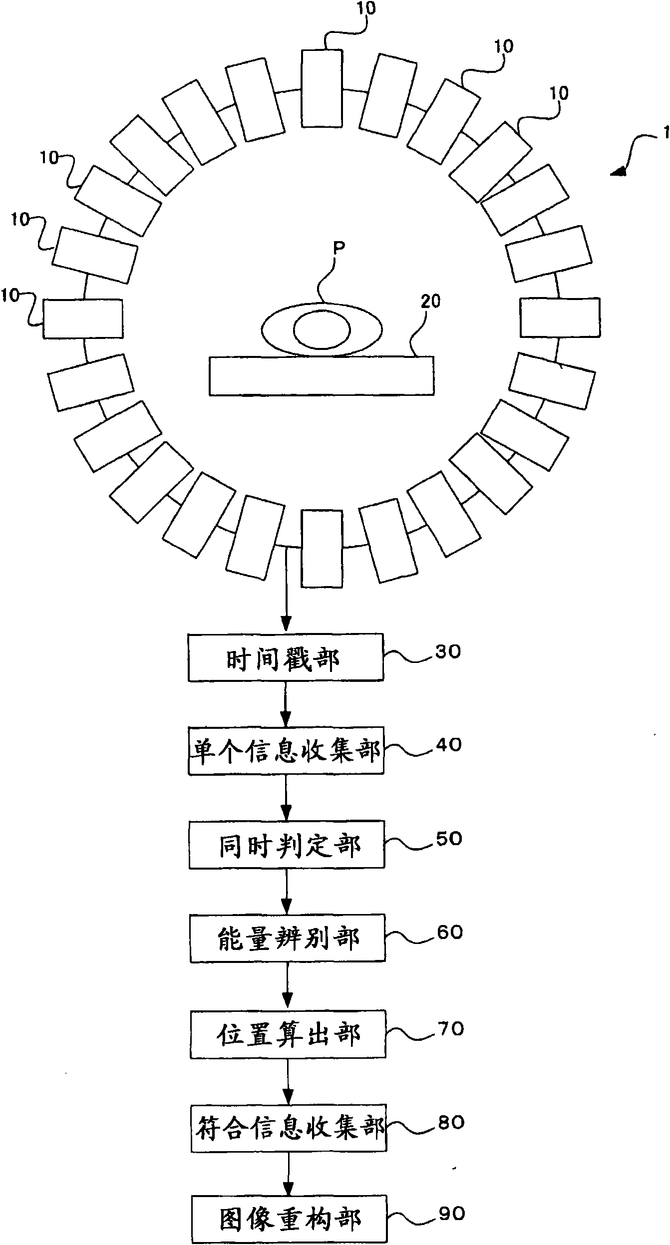
[0032] A PET apparatus and a method of generating a nuclear medicine image according to an embodiment of the present invention will be described with reference to the drawings.

[0033] image 3 The PET apparatus 1 shown detects gamma rays emitted from radioactive isotopes in a subject P. As shown in FIG. The PET apparatus 1 generates an image of the inside of the subject P based on the detection result.

[0034] As the radioisotope, positron radionuclides such as fluorodeoxyglucose can be used.

[0035] When the positrons emitted from the positron radionuclide combine with nearby free electrons to disappear, 511keV gamma rays are emitted in opposite directions (180° opposite directions) to each other. The PET apparatus 1 detects the gamma rays. The PET apparatus 1 determines the incident direction of the gamma ray by distinguishing the gamma ray incident at the same timing. The PET apparatus 1 assumes that radioactive isotopes exist on the trajectory of the incident direct...


PUM

PropertyMeasurementUnit
Thicknessaaaaaaaaaa

Abstract

The invention provides a positron emission tomography apparatus and a nuclear medical image generating method. In a case that a gamma ray has entered into a plurality of scintillators adjacent to each other simultaneously, a detector detects the gamma ray having entered simultaneously. A position calculator calculates the ratio of wave heights representing the energies of the detected gamma ray. The position calculator obtains a trajectory of such a gamma ray that a ratio of distances passed by the gamma ray inside the plurality of scintillators, respectively, coincides with the ratio of the wave heights. The position calculator obtains an intersection between the boundary of the plurality of scintillators and the trajectory, as a passing position of the gamma ray. A reconstructing part executes a back projection process with the trajectory passing through the calculated passing position as a projection position.

Description

technical field [0001] The present invention relates to a Positron Emission Tomography Apparatus (PET apparatus) for detecting gamma rays emitted from radioactive isotopes in a subject and a nuclear medicine image generation method. Background technique [0002] A PET device detects gamma (γ) rays emitted from radioactive isotopes in a subject, and generates an image (nuclear medicine image) based on the detection result. In the examination using the PET apparatus, a positron radionuclide (Positronradionuclide) such as fluorodeoxyglucose (FDG) as a radioactive isotope is supplied to the subject. A positron radionuclide is used as a tracer to label biological substances and the like in a subject. Physiological or biochemical functions are obtained from the concentration distribution of radioactive isotopes as data. This data is acquired as two-dimensional image data or three-dimensional image data. [0003] When positrons combine with free electrons to disappear, they emit...

Claims


Application Information

Patent Timeline
IPC IPC(8): A61B6/03G01T1/161
CPCG01T1/2985A61B6/037
Inventor 高山卓三勅使川原学梅原隆哉小森智康
Owner KK TOSHIBA
Learn More