Method for shear wave field formation and ultrasonic image system

An imaging method and technology of a shear wave subsystem, applied in the field of tissue imaging methods and systems based on shear waves

Active Publication Date: 2011-04-20
SHENZHEN MINDRAY BIO MEDICAL ELECTRONICS CO LTD
View PDF3 Cites 44 Cited by
  • Summary
  • Abstract
  • Description
  • Claims
  • Application Information

AI Technical Summary

Problems solved by technology

However, the method of manual palpation can only be limited to the tissues that can be touched by hand

Method used


Image

  • Method for shear wave field formation and ultrasonic image system
  • Method for shear wave field formation and ultrasonic image system
  • Method for shear wave field formation and ultrasonic image system

Examples


Embodiment Construction

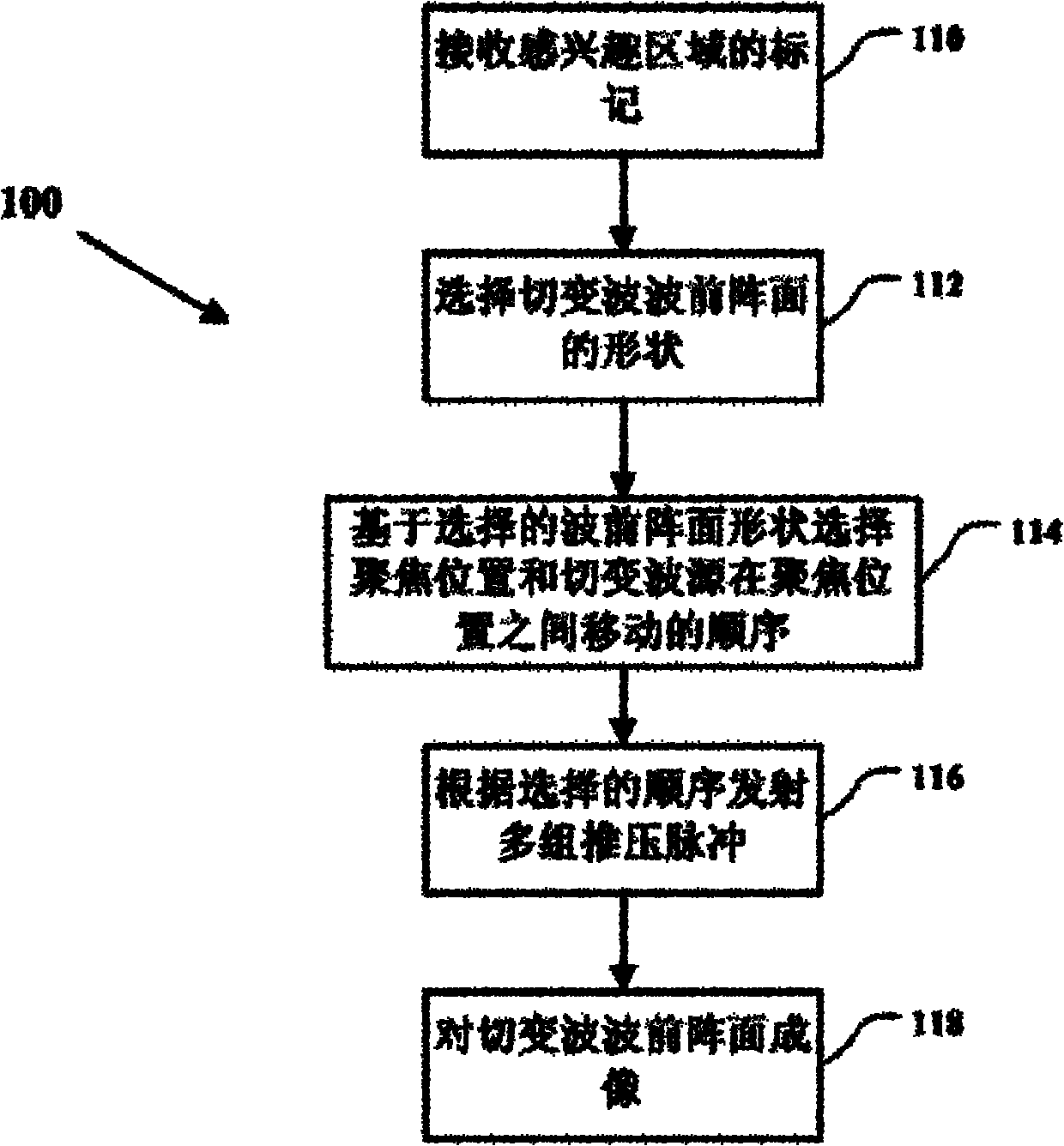
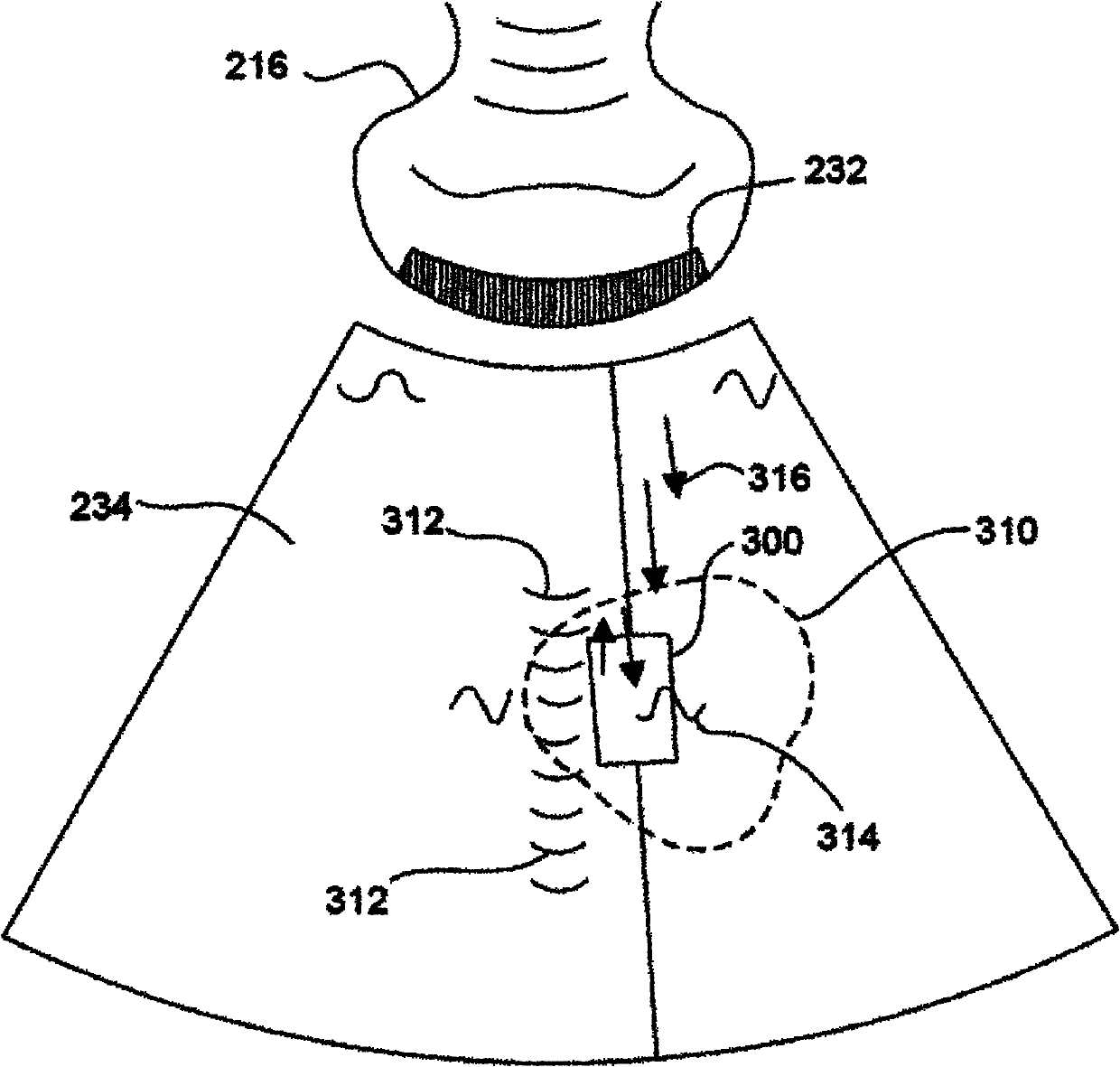
[0015] In the ultrasonic shear wave tissue imaging of the embodiment of the present invention, ultrasonic waves called "push pulses" are emitted, and these push pulses are focused inside the biological tissue. The pushing pulse generates a shear wave through the acoustic force inside the tissue, and the propagating direction of the shear wave is transverse to the propagating direction of the pushing pulse. The focal point of the push pulse inside the tissue may be referred to herein as a "shear wave source". Shear waves cause measurable time-varying displacements that are related to the viscoelasticity of the current tissue.

[0016] A larger shear wave intensity can cause a larger displacement of the tissue. Therefore, to increase the strength of the shear wave, a "supersonic" shear wave tissue imaging technique uses multiple sets of push pulses. Multiple sets of push pulses are delivered to focus on a closely spaced series of locations within the tissue. Thus, the shear w...


PUM


Abstract

The invention embodiment discloses a method for shear wave field formation and an ultrasonic image system. The system receives an indication of a region of interest and selects a shear wave front shape. The system also selects, based on the selected shear wave front shape, focus locations for a plurality of push pulses and a sequence for moving a shear wave source among the focus locations. The system transmits a series of push pulses according to the selected sequence, and determines a speed of the shear wave front as it passes through the region of interest. Changes in the speed of the shear wave front are related to changes in stiffness within the tissue.

Description

technical field [0001] The invention relates to the field of medical ultrasound imaging, in particular to a shear wave-based tissue imaging method and system thereof. Background technique [0002] Determining the relative stiffness of tissues can be used to diagnose disease and monitor treatment of disease. Conventional imaging techniques, such as conventional B-mode ultrasound imaging, do not clearly image sclerotic lesions or tissues. This sclerotic lesion or tissue can be diagnosed by manual palpation. However, the method of manual palpation can only be limited to the tissues that can be touched by hands, but it is powerless for the tissues that cannot be touched by hands in the deeper part of the body. Therefore, in the field of ultrasound imaging, it is necessary to develop a technique capable of imaging the viscoelasticity or hardness of biological tissue. Contents of the invention [0003] The embodiment of the present invention discloses an ultrasonic transverse...

Claims


Application Information

Patent Timeline
IPC IPC(8): A61B8/08
CPCG01S7/52036G01S7/52042A61B8/0833A61B5/0048A61B8/485A61B8/08
Inventor 姚林鑫
Owner SHENZHEN MINDRAY BIO MEDICAL ELECTRONICS CO LTD
Why Eureka
Try Eureka