Method for automatic computerized segmentation and analysis on thickness uniformity of intima media of carotid artery blood wall in sonographic image

A technology of vascular ultrasound and uniform thickness, which is applied in the directions of ultrasound/sonic/infrasonic image/data processing, calculation, image enhancement, etc., and can solve the problem of time-consuming

Inactive Publication Date: 2011-08-24
武汉上上智科技发展有限公司
View PDF7 Cites 32 Cited by
  • Summary
  • Abstract
  • Description
  • Claims
  • Application Information

AI Technical Summary

Problems solved by technology

The doctor's judgment of the intima-media boundary in these measurement activities directly depends on his training, subjective understanding of the boundary, experience, etc. Therefore, different doctors judge the intima-media boundary differently, and even the same doctor obtains the intima-media boundary at different times. The results are not completely consistent, in addition, it also has the disadvantage of taking a long time
Among the published patents, two

Method used


Image

  • Method for automatic computerized segmentation and analysis on thickness uniformity of intima media of carotid artery blood wall in sonographic image
  • Method for automatic computerized segmentation and analysis on thickness uniformity of intima media of carotid artery blood wall in sonographic image
  • Method for automatic computerized segmentation and analysis on thickness uniformity of intima media of carotid artery blood wall in sonographic image

Examples


Embodiment 1

[0064] A method for computer automatic segmentation and thickness uniformity analysis of carotid artery intima-media in vascular ultrasound images, characterized in that it includes the following steps, such as figure 2 Shown:

[0065] 1. Load an acquired original vascular ultrasound image into the computer connected to the ultrasound imager. If the pixel size of the vascular ultrasound image is different, the computer will automatically adjust the specific parameters according to the pixel size of the image (automatically obtained from the DICOM format) ;

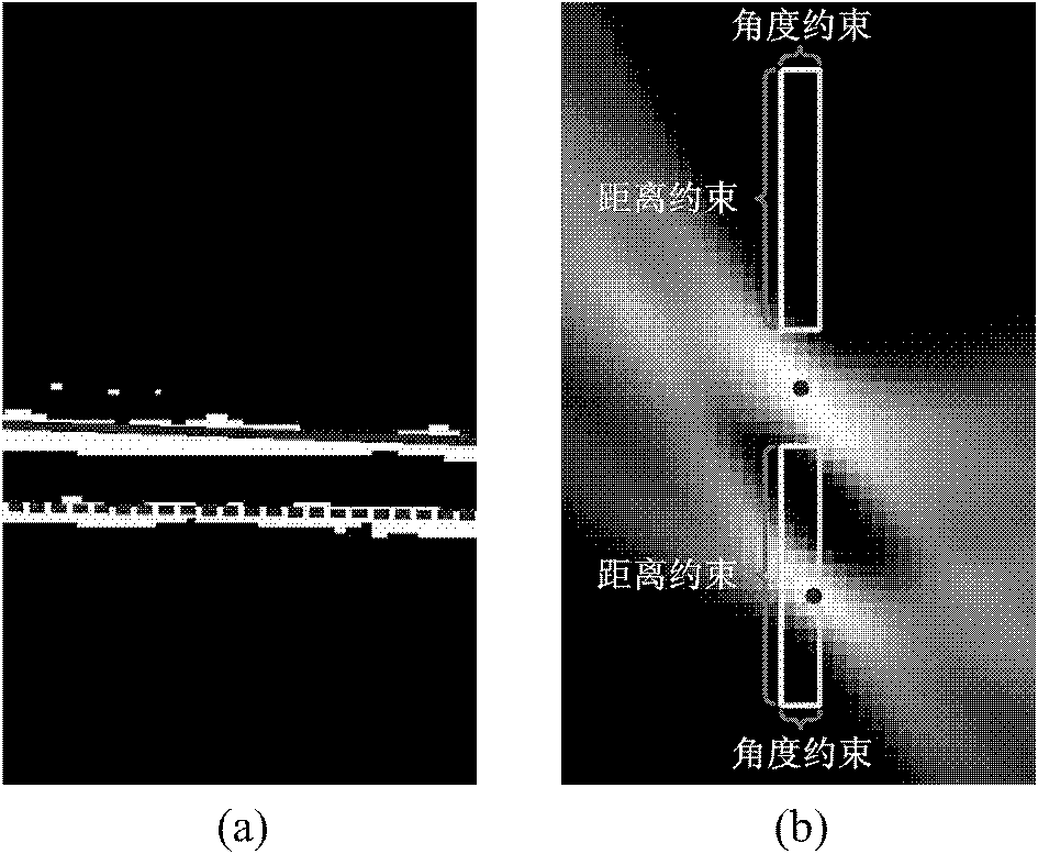
[0066] 2. Manually select a rectangular region of interest (ROI) containing the intima-media of the distal wall of the vessel, see Figure 6 Image in (a);

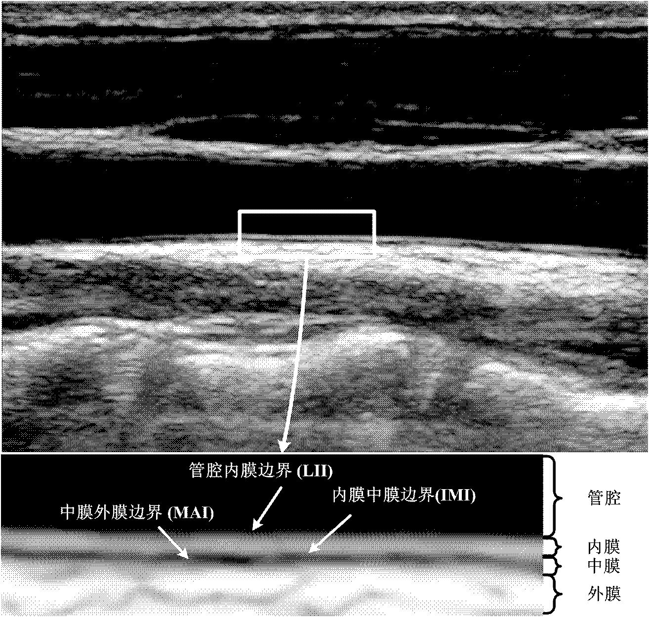
[0067] 3. Detect the evolution in the rectangular region of interest to obtain the precise contour lines of the two boundaries of LII (vascular lumen and intima boundary) and MAI (media and adventitia boundary) of the intima:

[0068] 3.1 Automatically detect the i...

Embodiment 2

[0108] According to the method of embodiment 1, to Figure 7 The image in (a) is automatically segmented to obtain the precise contour lines of the two boundaries of carotid artery intima-media LII and MAI, as shown in figure (b). Figure (c) is the artificially drawn contour lines of the two boundaries of carotid artery intima-media LII and MAI. According to the two boundary contour lines of LII and MAI of the intima-media of the carotid artery obtained by precise segmentation, the mean, standard deviation, coefficient of variation, percentage composition ratio and percentile of the thickness of each point of the intima-media in a section of blood vessel were counted. The results are shown in Table 1. This result shows that the present invention can segment the healthy intima-media (thicker) very well.

Embodiment 3

[0110] According to the method of embodiment 1, to Figure 8 The image in (a) is automatically segmented to obtain the precise contour lines of the two boundaries of carotid artery intima-media LII and MAI, as shown in figure (b). Figure (c) is the artificially drawn contour lines of the two boundaries of carotid artery intima-media LII and MAI. According to the two boundary contour lines of LII and MAI of the intima-media of the carotid artery obtained by precise segmentation, the mean, standard deviation, coefficient of variation, percentage composition ratio and percentile of the thickness of each point of the intima-media in a section of blood vessel were counted. The results are shown in Table 1. This result shows that the present invention can correctly segment the intima-media thickened but not yet lesioned (thickness less than 1 mm).


PUM

PropertyMeasurementUnit
Thicknessaaaaaaaaaa

Abstract

The invention belongs to the field of intersection of computer technologies and medical images, and particularly relates to a method for automatic computerized segmentation and analysis on the thickness uniformity of an intima media of a carotid artery blood wall in a sonographic image. The method comprises the following steps of: loading an original blood wall sonographic image; selecting a rectangular region of interest (ROI) containing carotid artery intima media of a far side blood wall; artificially defining or automatically detecting an original contour line by a computer; calculating aone-way edge map; performing binarization and segmentation; detecting two straight segments PLi and SLi serving as partial and correction connection of a lumen intima interface (LII) and a media adventitia interface (MAI) on a sub-image through Hough transformation; obtaining an intima media boundary contour line by adopting double-Snake model evolution; and calculating the thickness uniformity on the basis of two boundaries obtained from segmentation. By the method, problems of noise and discontinuous boundaries in the images can be effectively solved, the purpose of accurate segmentation ofthe intima media boundary is fulfilled and workloads of a doctor can be reduced. Moreover, thickness uniformity parameters obtained on the basis of the method can provide more information for early analysis on atherosclerosis.

Description

technical field [0001] The invention belongs to the intersecting field of computer technology and medical images, and in particular relates to a computer automatic segmentation and thickness uniformity analysis method of carotid artery intima-media in vascular ultrasound images. Background technique [0002] Ischemic cardiovascular and cerebrovascular disease is a major disease that endangers human health, and its main pathological basis is atherosclerosis. Carotid artery intima-media thickness (Intima Media Thickness, IMT) reflects the status of atherosclerosis, and it is highly correlated with the incidence of myocardial infarction and stroke. Therefore, it has been used to predict the risk of cardiovascular and cerebrovascular diseases in the past two decades. One of the important indicators. [0003] Carotid artery intima-media thickness was measured on ultrasound images of vessels. The normal vascular intima-media appears as a hyperechoic and hypoechoic two-layer band...

Claims


Application Information

Patent Timeline
IPC IPC(8): G06T5/00G06F19/00A61B8/08
Inventor 程新耀宋恩民周渊
Owner 武汉上上智科技发展有限公司
Why Eureka
Try Eureka