Contrast-enhanced ultrasound assessment of liver blood flow for monitoring liver therapy

A contrast-enhanced, liver-based technology, applied in the field of medical imaging systems, can solve the problems of invisibility, shrinkage, and inability to perfectly reproduce the exact same scanning plane and ROI position

Inactive Publication Date: 2012-07-11
KONINKLIJKE PHILIPS ELECTRONICS NV
View PDF14 Cites 4 Cited by
  • Summary
  • Abstract
  • Description
  • Claims
  • Application Information

AI Technical Summary

Problems solved by technology

Moreover, such protocols assume that the response (or lack of response) to a single tumor treatment represents the systemic liver response to treatment, which may also be inaccurate
[0009] When using the above protocol in the context of oncology therapy monitoring, it is extremely challenging to compare parameter values ​​obtained at different time points dur

Method used


Image

  • Contrast-enhanced ultrasound assessment of liver blood flow for monitoring liver therapy
  • Contrast-enhanced ultrasound assessment of liver blood flow for monitoring liver therapy
  • Contrast-enhanced ultrasound assessment of liver blood flow for monitoring liver therapy

Examples


Embodiment Construction

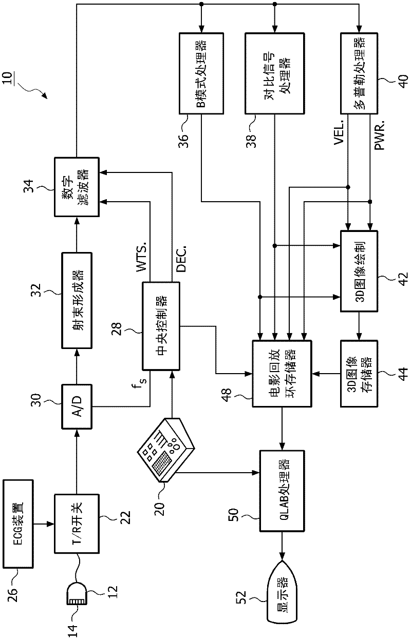
[0034] The invention can be implemented in an imaging and / or evaluation system having a real-time low mechanical index CEUS imaging modality to obtain contrast-enhanced images. Because the illustrated system incorporates a low mechanical index, rupture of contrast agent microbubbles can be minimized or completely prevented, allowing accurate imaging, observation, and / or Quantify.

[0035] Embodiments of the present invention do not focus on a single liver target lesion, but instead can image and quantify blood flow from the hepatic aorta (hereinafter referred to as hepatic artery) and main portal vein (hereinafter referred to as portal vein) to the liver to assess Systemic response of the liver to disposition. US 2009 / 0124907A1 (Bruce et al.), published May 14, 2009, entitled "Ultrasonic Diagnostic Imaging System and Method for Detecting Lesion of the Liver", describes how contrast flow in the hepatic artery and portal vein can be exploited, based on For example, the time of...


PUM


Abstract

A method for assessing a liver includes acquiring image information including contrast-enhanced ultrasound images of the liver. A location of the main hepatic artery (MHA) and a location of the main portal vein (MPV) of the liver are identified in at least one of the contrast-enhanced ultrasound images of the liver. Time-intensity information corresponding to perfusion of a contrast agent in the MHA and the MPV is obtained. A biomarker index value (BIV) which is a function of the time-intensity information corresponding to the perfusion of contrast agent in the MHA and the time-intensity information corresponding to the perfusion of contrast agent in the MPV is determined.

Description

[0001] This application claims the benefit of US Provisional Application No. 61 / 247655, filed October 1, 2009, which is hereby incorporated by reference. technical field [0002] The present system relates to medical imaging systems, and more particularly to systems for assessing liver blood flow to monitor liver therapy. Background technique [0003] Currently, monitoring liver therapy with contrast-enhanced ultrasound (CEUS) is performed by identifying a single target liver lesion (tumor) and quantifying its blood flow and fractional blood volume. Such protocols assume that response (or lack of response) to treatment of a single target lesion represents a systemic response to treatment, but this is not a valid assumption in all cases. [0004] Accurate early assessment of treatment response is key to effective management of cancer patients and evaluation of new therapeutic compounds. Histological determination of mean intratumoral microvessel density (MVD) is the most com...

Claims


Application Information

Patent Timeline
IPC IPC(8): A61B8/00A61B8/06G06T7/00
CPCG06T7/0016G06T2207/10132A61B8/5223A61B8/469A61B8/481A61B8/06G06T2207/30056G16H50/30
Inventor T·戈捷E·L·S·利恩
Owner KONINKLIJKE PHILIPS ELECTRONICS NV
Why Eureka
Try Eureka