Evaluation of carotid plaque using contrast-enhanced ultrasound imaging

A technology of contrast enhancement and ultrasound imaging, which can be used in image enhancement, ultrasound/sonic/infrasound diagnosis, applications, etc., and can solve problems such as cerebral vascular obstruction and stroke

Active Publication Date: 2019-10-01
KONINKLJIJKE PHILIPS NV
View PDF10 Cites 0 Cited by
  • Summary
  • Abstract
  • Description
  • Claims
  • Application Information

AI Technical Summary

Problems solved by technology

Since the carotid artery provides blood supply to the brain, certain substances in the bloodstream of the carotid artery can cause blockage of the blood vessel in the brain and a potential risk of stroke

Method used


Image

  • Evaluation of carotid plaque using contrast-enhanced ultrasound imaging
  • Evaluation of carotid plaque using contrast-enhanced ultrasound imaging
  • Evaluation of carotid plaque using contrast-enhanced ultrasound imaging

Examples


Embodiment Construction

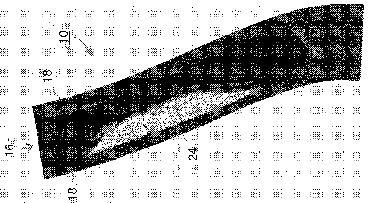
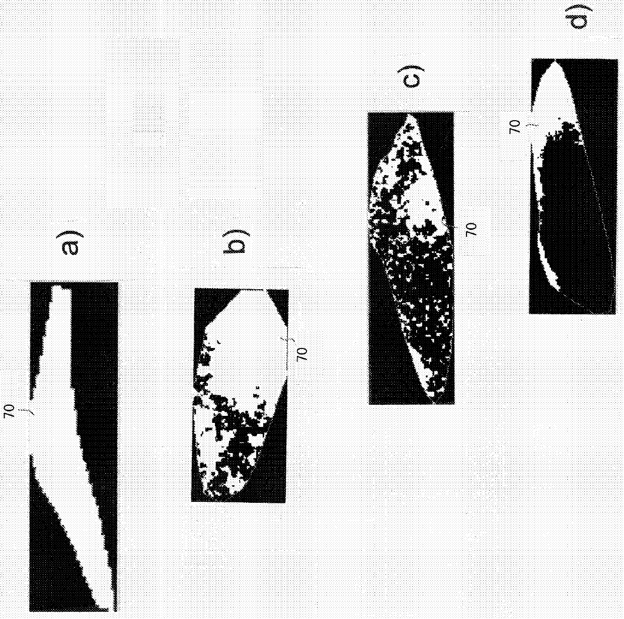
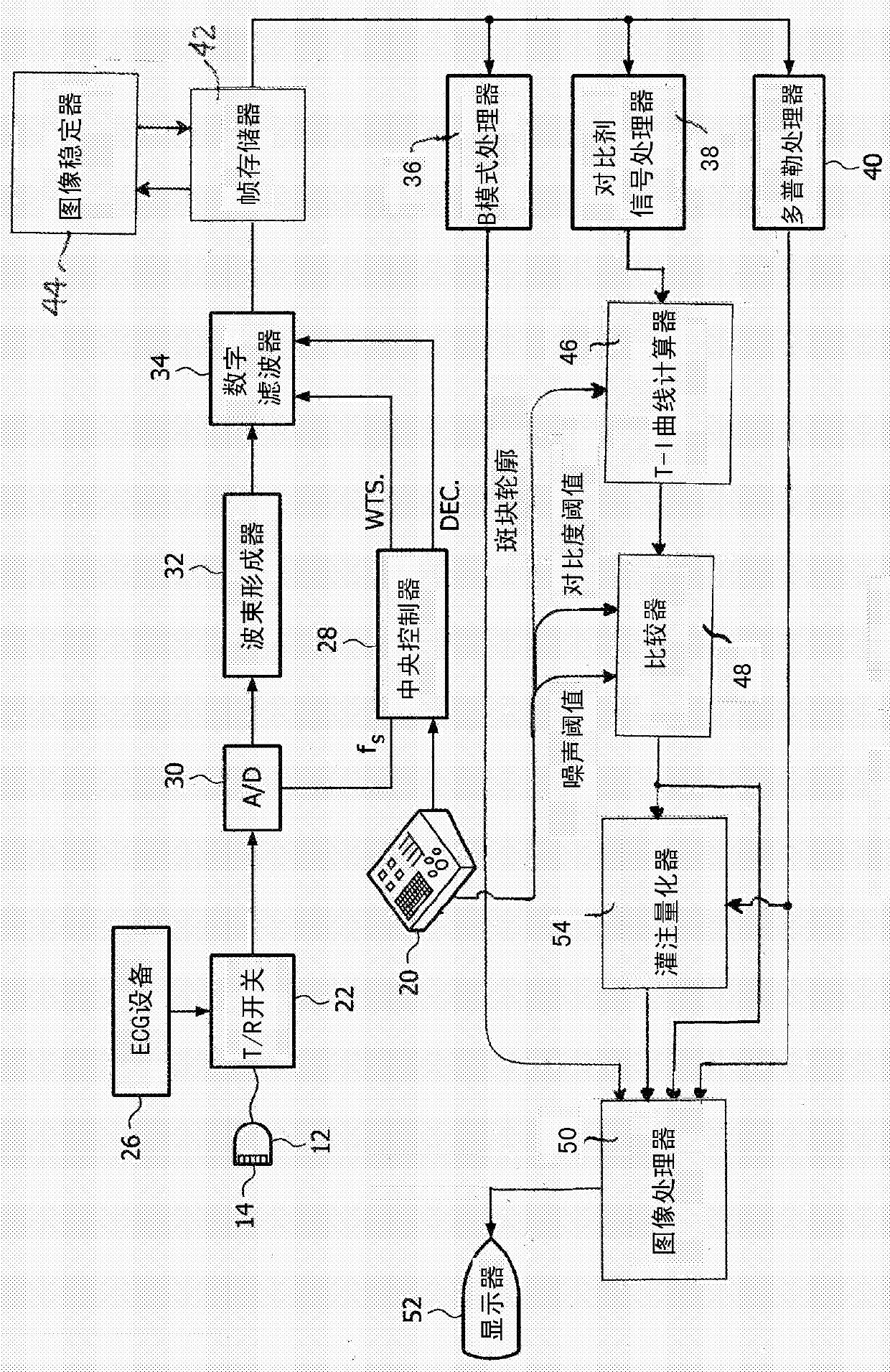
[0015] In some embodiments, the present invention provides an ultrasound imaging system, eg, an ultrasound diagnostic imaging system, for the assessment of plaque by contrast enhanced ultrasound. The system can include an ultrasound imaging probe with an array transducer that acquires a sequence of ultrasound images of the plaque during contrast agent delivery. In certain embodiments, the system can be configured to include a processor, memory, and other structure capable of acting as a time-intensity curve calculator that forms The time-intensity curve of , and a comparator that identifies pixels in the plaque image where perfusion is present. In one embodiment, the comparator is further operable to compare pixel intensity values ​​before and after the arrival of the contrast agent in the plaque. The system can also include a display that shows the degree of perfusion in the plaque. In certain embodiments, the system can include an image processor that generates an anatomic...


PUM


Abstract

An ultrasound system and method are described for acquiring a sequence of ultrasound images of a carotid artery during delivery of a contrast agent. Plaques in the image shown are identified and time-intensity curves are calculated for pixels in the image. Intensity values ​​before and after arrival of the contrast agent are compared to identify pixels or groups of pixels with perfusion. An anatomical image can be created showing the intensity of perfusion and the area of ​​the image of the plaque present, or the perfusion can be quantified by determining the percentage of pixels in the plaque exhibiting perfusion. The extent and degree of perfusion is an indicator of the risk of plaque particles in the bloodstream that may cause stroke-related symptoms.

Description

technical field [0001] The present invention relates to ultrasound diagnostic imaging systems, and in particular to the use of contrast-enhanced ultrasound imaging to assess stroke risk. Background technique [0002] The development of plaque in the vasculature, especially atherosclerosis in the carotid arteries, occurs frequently in many patients. Plaque can build up to a point where partial and even complete blockage of blood vessels can occur. figure 1 A blood vessel 110 is illustrated with the vessel wall 18 partially cut away to show the interior lumen 16 in which plaque accumulation 24 is visible. Since plaque 24 has filled most of the lumen at its thickest point, it has partially blocked blood flow through the vessel. Plaque not only restricts the delivery of fresh blood to tissues and organs downstream of the blockage, but also traps blood clots in the narrowed lumen, blocking all blood flow. Additional events of equal severity could be caused by carotid plaque. ...

Claims


Application Information

Patent Timeline
Patent Type & Authority Patents(China)
IPC IPC(8): G06T7/00
CPCG06T2207/10132G06T2207/10136G06T2207/30101G06T2207/30104G06T7/0016A61B8/06A61B8/4444A61B8/5223H04N23/683G06F18/22A61B8/4494A61B8/461A61B8/481G06T7/20G06T2207/20021
Inventor M·阿韦基乌D·克里斯托菲迪斯E·L·S·利恩
Owner KONINKLJIJKE PHILIPS NV
Why Eureka
Try Eureka