Method for quantifying drug delivery using contrast-enhanced ultrasound
a contrast-enhanced ultrasound and drug delivery technology, applied in ultrasonic/sonic/infrasonic image/data processing, tomography, applications, etc., can solve the problem of inability to provide fix-quantity drug delivery, inferior sensibility of partial contrast agent, additional challenges, etc. problem, to achieve the effect of improving the signal intensity, high mechanical index, and specific mechanical index of ultrasound
- Summary
- Abstract
- Description
- Claims
- Application Information
AI Technical Summary
Benefits of technology
Problems solved by technology
Method used
Image

Examples
embodiments
Embodiment 1
An Ultrasound Quantifying System for Monitoring Drug Delivery
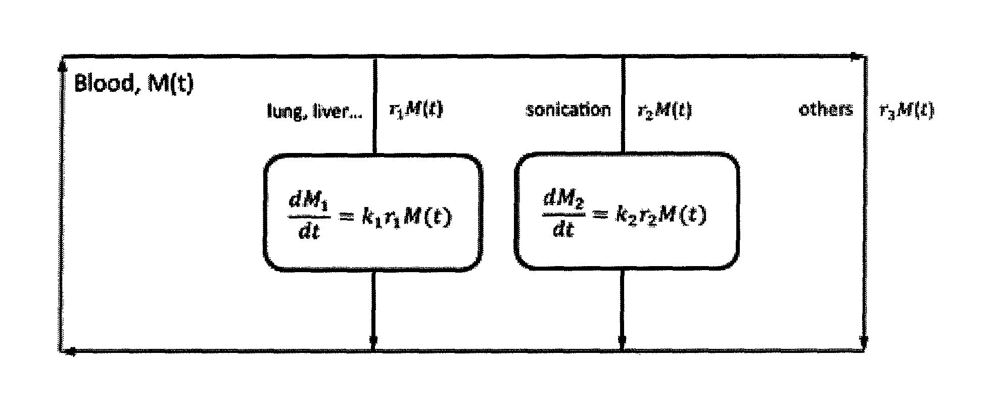
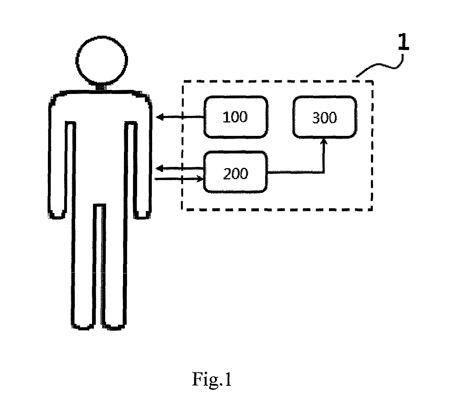
[0026]The ultrasound quantifying system (1) for monitoring drug delivery uses a contrast-enhanced ultrasound system to measure the signal intensity of peripheral vessels in order to quantify the drug delivery at target site. FIG. 1 shows the basic structure of the ultrasound quantifying system (1) of the presentation. Wherein the ultrasound quantifying system (1) includes: an injection device (100) that injects microbubble contrast agent into the tested object via a catheter or an intravenous injection; an ultrasound device (200) that records the variation of ultrasound signal intensity of the peripheral vessels of the tested object; and a signal processing device (300) that processes the ultrasound signal data obtained from the peripheral vessels of the tested object, converts the data into systemic microbubble concentration and thus calculates the drug delivery at the target site.
[0027]The ultrasound device (...
embodiment 2
Ultrasound Signal Saturation Experiment
[0031]The rats averaging 330 g were anesthetized intraperitoneally with chloral hydrate of 450 mg / kg. Then the rats were placed in a supine position and the hairs of both things were shaved for ultrasound experiment. A 24-gauge catheter was placed in the tail vein and a three-way stopcock was attached to the end of the catheter with one branch used to administer microbubble contrast agents in the vein bolus and the second for flushing with physiologic saline. The microbubble contrast agents can be anyone of the following substances: ALBUNEX®, SONOZOID®, SONOVUE®, SONOVIST®, OPTISON®, LEVOVIST® or DEFINITY®. In this preferred embodiment, the powder contrast agent SonoVue (Bracco, Milan, Italy) was used in this study. 5 mL of physiologic saline was added to 25 mg of the powder in order to make a microbubble contrast agent suspension of sulfur hexafluoride with the average diameter of 2.5 μm which is considered to be of no difference with saline i...
embodiment 3
Microbubble Destruction Experiment
[0035]In this preferred embodiment, two sets of ultrasound equipments are used. Besides the above-mentioned Philips iU22 ultrasound system (210), another Philips iU22 ultrasound system equipped with a L17-5 linear array transducer (220, frequency range 5˜17 MHz) was used for microbubbles destruction. Wherein, for the first ultrasound system (210), the probe of L12-5 linear array transducer still held orthogonal to the skin and parallel to the long axis of the thigh and the right femoral artery is still chosen for signal measurement. And, for the second ultrasound system (220), the probe of L17-5 linear array transducer was held orthogonal to and in full contact with the skin of the left upper thigh and parallel to the long axis of the thigh. The ultrasound was focused on the adductor muscles of the left hind limb. The transducer position and contact area were the same for each experiment in this preferred embodiment. The experimental setup is shown ...
PUM
 Abstract
Description
Claims
Application Information
 - R&D
- Intellectual Property
- Life Sciences
- Materials
- Tech Scout
- Unparalleled Data Quality
- Higher Quality Content
- 60% Fewer Hallucinations
Browse by: Latest US Patents, China's latest patents, Technical Efficacy Thesaurus, Application Domain, Technology Topic, Popular Technical Reports.
© 2025 PatSnap. All rights reserved.Legal|Privacy policy|Modern Slavery Act Transparency Statement|Sitemap|About US| Contact US: help@patsnap.com