Method of diagnosis

a diagnostic method and a technology of stefin a, applied in the field of diagnostic methods, can solve the problems of inconclusive clinical management of dcis patients, and inability to accurately predict which patients will develop invasive cancer, etc., to achieve the effect of increasing the activity of cathepsin protease, increasing the risk of neoplasia progression, and decreasing the expression of stefin

Pending Publication Date: 2019-01-31
LA TROBE UNIVERSITY
View PDF0 Cites 4 Cited by
  • Summary
  • Abstract
  • Description
  • Claims
  • Application Information

AI Technical Summary

Benefits of technology

[0074]Preferably, in the level of expression of Stefin A is assessed by detecting RNA transcripts, cDNA transcribed from the RNA transcripts and / or a protein expression product from the RNA transcripts and / or by detecting cathepsin protease activity, in particular cathepsin B, wherein increased cathepsin protease activity is indicative of a decrease in the level of Stefin A expression and an increased risk of progression of the neoplasia.

Problems solved by technology

A malignant neoplasm manifests a greater degree of autonomy, is capable of invasion and metastatic spread, may be resistant to treatment, and may cause death.
Progression of DCIS to IDC significantly increases the risk of tumour cell dissemination and subsequent metastasis.
However it is currently not possible to accurately predict which patients will develop invasive cancer.
However, although DCIS represents a significant (up to 25%) fraction of newly diagnosed breast cancer cases, the clinical management of DCIS patients is still inconclusive.
In the absence of the ability to discriminate between DCIS with differing prognosis, current methods of treating these cancers continue to follow the long used protocol of surgical excision (if possible) followed by radiotherapy and / or chemotherapy These treatments are associated with severe side effects including disfigurement and scarring from surgery (e.g. mastectomy or limb amputation), severe nausea and vomiting from chemotherapy, and most significantly, damage to normal tissues such as the hair follicles, gut and bone marrow which is induced as a result of the relatively non-specific targeting mechanism of the toxic drugs which form part of most cancer treatments.
To the extent that a proportion of DCIS are unlikely to progress, patients are unnecessarily subjected to these aggressive forms of treatment.
Currently there is no means to reliably and routinely assess and / or monitor a patient in this way.
However, still more unexpectedly, it has been determined that patients who maintain normal Stefin A expression levels in the myoepithelial cells in fact need not undergo treatment at all, in particular radiotherapy.

Method used


Image

  • Method of diagnosis
  • Method of diagnosis
  • Method of diagnosis

Examples


example 1

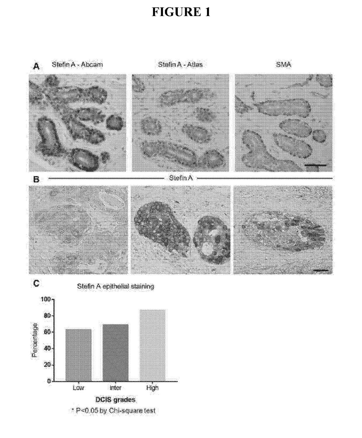
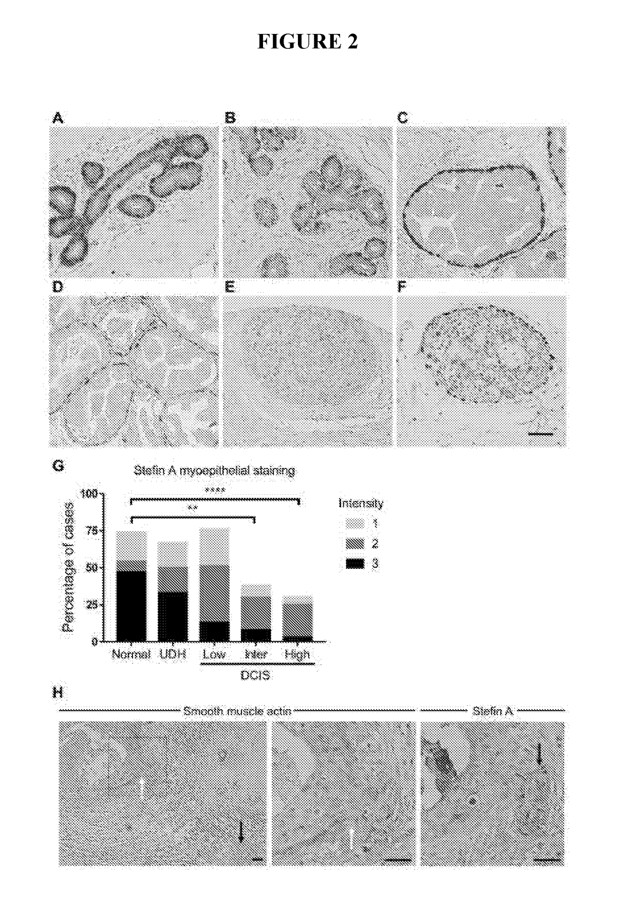
[0174]Analysis of Stefin A expression in a cohort of 200 patient samples spanning normal, hyperplasia and low, intermediate and high grade DCIS lesions has revealed that the expression of Stefin A is significantly reduced with increasing DCIS grade. The loss of Stefin A in high grade DCIS lesions, those more likely to progress to invasive carcinoma, indicates that myoepithelial Stefin A suppresses the DCIS to invasive carcinoma transition. In summary a 3D culture system has been used to test this, as described below

[0175]Method and Materials

Immunohistochemistry (IHC)

[0176]For human tissues, normal breast sections and primary breast carcinoma samples were obtained from Sandra O'Toole at the Royal Prince Alfred Hospital (RPAH) either as full-faced slides (for the micro-invasive carcinoma) or in a tissue microarray (TMA) (Zardawi et al.). The use of archived human tissues was approved by the HREC of RPAH (approval number X15-0388 (SSA / 16 / RPAH / 397)). Sections (formalin-fixed, paraffin e...

example 2

Recurrence of Invasive Cancer

[0214]Samples were surgically resected from pre-invasive lesions (DCIS) from 3 patients in 1997, 2004 and 2001 respectively (Identified as Patient 1997, Patient 2004 and Patient 2001) and the samples were archived. These patients were subsequently followed up clinically over 10 years.

[0215]The archived samples of Patient 1997, Patient 2004 and Patient 2001 were subsequently analysed for Stefin A (FIG. 14). Staining of Stefin A was carried out as previously described (Immunohistochemistry (IHC)). Patient 1997 and 2004 showed low to no staining of Stefin A and later developed a clinically diagnosed invasive neoplastic lesion, confirmed by methods such as mammogram and core biopsy.

[0216]In contrast, the archived sample of Patient 2001 displays normal levels of Stefin A. Patient 2001 has had annual follow up for approximately 10 years and has no recurrence of breast cancer.


PUM

PropertyMeasurementUnit
temperatureaaaaaaaaaa
pHaaaaaaaaaa
pHaaaaaaaaaa

Abstract

The present invention relates generally to a method of detecting a risk of the progression of a pre-invasive neoplasia of the glandular epithelium. More particularly, the present invention provides a method of detecting a risk of the progression from a pre-invasive breast neoplasia by screening for the level of expression of Stefin A in the myoepithelial cells. The method of the present invention is useful in a range of applications including, but not limited to, assessing a neoplastic condition, monitoring the progression of such a condition, predicting the likelihood of a subject progressing to a more advance disease state or informing decisions in relation to the design of treatment schedules.

Description

FIELD OF THE INVENTION[0001]The present invention relates generally to a method of detecting a risk of the progression from a pre-invasive neoplasia of the glandular epithelium. More particularly, the present invention provides a method of detecting a risk of the progression from a pre-invasive breast neoplasia by screening for the level of expression of Stefin A in the myoepithelial cells. The method of the present invention is useful in a range of applications including, but not limited to, assessing a neoplastic condition, monitoring the progression of such a condition, predicting the likelihood of a subject progressing to a more advance disease state or informing decisions in relation to the design of treatment schedules.BACKGROUND OF THE INVENTION[0002]The reference in this specification to any prior publication (or information derived from it), or to any matter which is known, is not, and should not be taken as an acknowledgment or admission or any form of suggestion that that...

Claims


Application Information

Patent Timeline
Patent Type & Authority Applications(United States)
IPC IPC(8): G01N33/574A61N5/00A61K39/00
CPCG01N33/57415A61N5/00A61K39/0011G01N2333/8139C12Q1/68C12Q1/6886C12Q2600/118C12Q2600/158
Inventor PARKER, BELINDA SHEREEDUIVENVOORDEN, HENDRIKA MARTHA
Owner LA TROBE UNIVERSITY
Why Eureka
Try Eureka