Contrast-enhanced ultrasound imaging method and system

A technology of contrast-enhanced ultrasound and imaging method, which is applied in the field of ultrasound, which can solve the problems of different contrast effects, poor imaging effects of contrast agents, and the inability of ultrasonic equipment to meet different contrast imaging in different regions, so as to achieve accurate imaging effects and a wide range of applications.

Active Publication Date: 2019-01-25
SONOSCAPE MEDICAL CORP
View PDF6 Cites 0 Cited by
  • Summary
  • Abstract
  • Description
  • Claims
  • Application Information

AI Technical Summary

Problems solved by technology

As a result, different contrast agents have different contrast effects under the same contrast imaging conditions, and the current ultrasound equipment basically only has the same set of parameters

Method used


Image

  • Contrast-enhanced ultrasound imaging method and system
  • Contrast-enhanced ultrasound imaging method and system
  • Contrast-enhanced ultrasound imaging method and system

Examples


Embodiment Construction

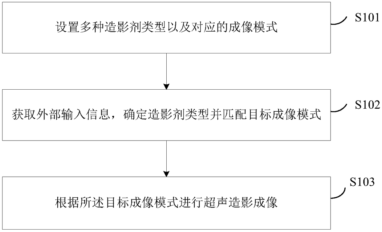
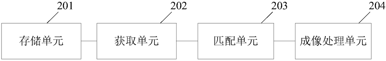
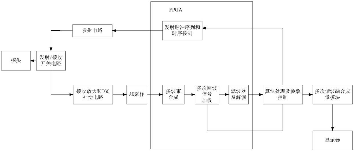
[0035] The following will clearly and completely describe the technical solutions in the embodiments of the present invention with reference to the accompanying drawings in the embodiments of the present invention. Obviously, the described embodiments are only some, not all, embodiments of the present invention. Based on the embodiments of the present invention, all other embodiments obtained by persons of ordinary skill in the art without making creative efforts belong to the protection scope of the present invention.

[0036] The terms "first" and "second" in the specification and claims of the present invention and the above drawings are used to distinguish different objects, rather than to describe a specific order. Furthermore, the terms "comprising" and "having", and any variations thereof, are intended to cover a non-exclusive inclusion. For example, a process, method, system, product or apparatus comprising a series of steps or units is not defined by listed steps or u...


PUM


Abstract

The invention discloses a contrast-enhanced ultrasound imaging method and system, the method comprises: setting a plurality of contrast agent types and a corresponding imaging mode, the imaging mode comprises parameters, algorithms and/or programs adapted to the contrast agent types; acquiring external input information, determining a contrast agent type and matching a target imaging mode; contrast ultrasound imaging is performed according to the target imaging mode. Since the imaging mode corresponding to each contrast agent is adopted, the imaging of the contrast agent is more accurate, andthe ultrasound apparatus can be applied to different contrast agents in different countries, so that the application range is wider.

Description

technical field [0001] The present invention relates to the field of ultrasound technology, in particular to an ultrasound contrast imaging method and system. Background technique [0002] Ultrasound contrast agents are liquids containing gas bubbles a few microns in diameter. Utilizing the strong scattering properties of liquid containing air bubbles to ultrasonic waves, ultrasound contrast agents are injected clinically into human blood vessels, and the blood supply of tissue lesions is evaluated through contrast agent perfusion imaging, so as to serve the purpose of auxiliary diagnosis. [0003] At present, due to the different approvals of ultrasound contrast agents in various countries, the types of clinical contrast agents entering each country or region are different. For example, in mainland China, only one contrast agent, SonoVue, is allowed to be used clinically, and it can be supported in Japan. SonoVue and Sonozoid enter the imaging clinic. Due to the different...

Claims


Application Information

Patent Timeline
IPC IPC(8): A61B8/08
CPCA61B8/0891A61B8/467A61B8/481
Inventor 朱建武冯乃章
Owner SONOSCAPE MEDICAL CORP
Why Eureka
Try Eureka