Method for enhancing liver blood vessel and simultaneously dividing liver from blood vessel in CTA (computed tomography imaging) image

A technology in liver blood vessels and images, which is applied in the field of medical image processing and can solve problems such as difficulty in segmenting the liver

Active Publication Date: 2012-07-25
ZHEJIANG UNIV
View PDF3 Cites 17 Cited by
  • Summary
  • Abstract
  • Description
  • Claims
  • Application Information

AI Technical Summary

Problems solved by technology

In addition, since the abdominal scan image contains many irrelevant tissues, such as stomach, heart, spleen, etc., which are very close to the gray value of

Method used


Image

  • Method for enhancing liver blood vessel and simultaneously dividing liver from blood vessel in CTA (computed tomography imaging) image
  • Method for enhancing liver blood vessel and simultaneously dividing liver from blood vessel in CTA (computed tomography imaging) image
  • Method for enhancing liver blood vessel and simultaneously dividing liver from blood vessel in CTA (computed tomography imaging) image

Examples


Embodiment Construction

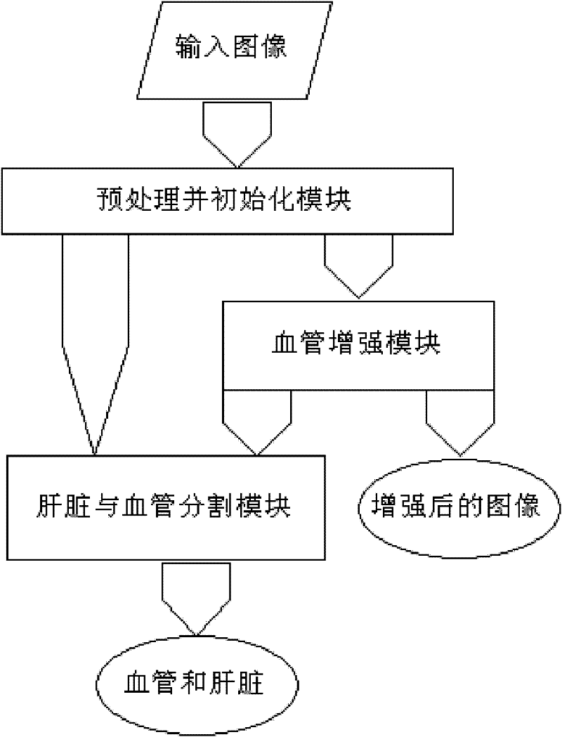
[0062] figure 1 The process of blood vessel enhancement and liver and blood vessel segmentation in the CTA scan image is shown in the figure. The specific process is as follows:

[0063] In the implementation process, the input of blood vessel and liver segmentation can be an image enhanced with blood vessels, or an image without enhancement, and the former is adopted in this embodiment.

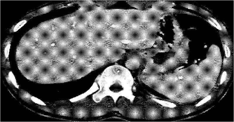
[0064] 1. Input liver CTA or MRA scan image I 1 , the size is 512×512×368, and the window width and level are adjusted so that the gray scale range of the liver and blood vessels is mainly between 0:255. figure 2 It is the 88th slice image of the three-dimensional liver data cross section. Perform Gaussian denoising on the image: I=I 1 *G δ , * is the convolution operator, is a Gaussian kernel function with window δ. In this example, δ=0.5. The initialization adopts interactive software, and randomly selects a liver area without blood vessels inside the liver.

[0065] 2. In an emb...


PUM


Abstract

The invention relates to processing of a medical image and in particular relates to a method for enhancing the liver blood vessel and simultaneously dividing the liver from the blood vessel in a CTA (computed tomography imaging) image. The method comprises the following steps of: preprocessing an image by means of gaussian convolution; computing the anisotropic characteristic value of each point of the image, and further conforming the anisotropic oval neighbourhood of each point; computing a grey level histogram in each neighbourhood, initiating a liver region, and computing a grey level histogram in the liver region; computing the wasserstein distance between the grey level histogram in each point neighbourhood and the grey level histogram in the liver region; enhancing the blood vessel according to the wasserstein distance and the anisotropic characteristic of the neighbourhood; and dividing the liver from the blood vessel. According to the method provided by the invention, the negative effects caused by the low contrast ratio, the noise, the fuzzy boundary and the like can be overcome, and the blood vessel identifying and dividing accuracy rate can be greatly improved, thus the anatomical structure information of the liver blood vessel can be exactly obtained.

Description

technical field [0001] The invention relates to the field of medical image processing, in particular to the enhancement of liver blood vessels in CT (computed tomography contrast enhancement) images and the simultaneous segmentation of liver entities and blood vessels. Background technique [0002] The structure of liver and blood vessels is very important for the diagnosis of liver disease and the guidance of liver disease surgery. CTA (computed tomography with contrast enhancement) is one of the most commonly used imaging diagnostic techniques for liver and blood vessels. With CTA, physicians can obtain a series of two-dimensional CT slices with enhanced blood vessels. Liver volume is an important reference index for liver surgery. However, abdominal CTA images often have unfavorable factors such as low contrast, high signal-to-noise ratio, blurred borders, and adhesion of the liver to other tissues with similar gray levels, making accurate liver segmentation a difficult ...

Claims


Application Information

Patent Timeline
IPC IPC(8): G06T5/00
Inventor 孔德兴彭佳林吴法王金伟
Owner ZHEJIANG UNIV
Why Eureka
Try Eureka