Implantable ophthalmic MEMS sensor devices and methods for eye surgery

An eye and implant technology, applied in the field of implantable ophthalmic microelectromechanical system sensing devices and eye surgery, can solve the problems of rapid IOP changes that cannot be detected and lost

Inactive Publication Date: 2012-10-03
ORTHOMEMS
View PDF7 Cites 9 Cited by
  • Summary
  • Abstract
  • Description
  • Claims
  • Application Information

AI Technical Summary

Problems solved by technology

Patients may undergo regular checkups that may not be ideal for quickly detecting scar tissue formation
Also, spikes in IOP and rapid changes in IOP that cause nerve damage may go undetected, so that, at least in some cases, patients may continue to lose at least some acuity of vision after surgery

Method used


Image

  • Implantable ophthalmic MEMS sensor devices and methods for eye surgery
  • Implantable ophthalmic MEMS sensor devices and methods for eye surgery
  • Implantable ophthalmic MEMS sensor devices and methods for eye surgery

Examples


Embodiment Construction

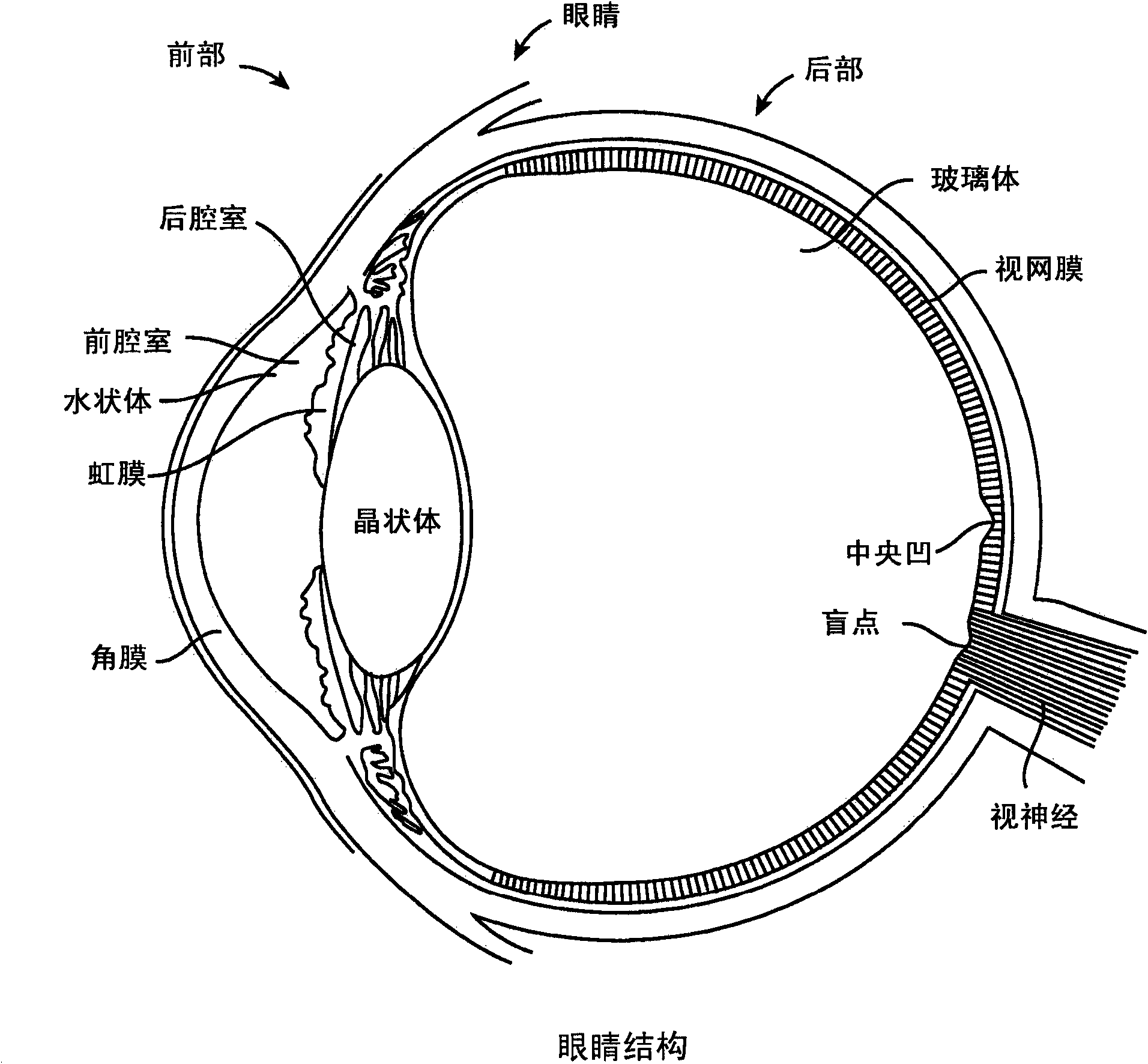
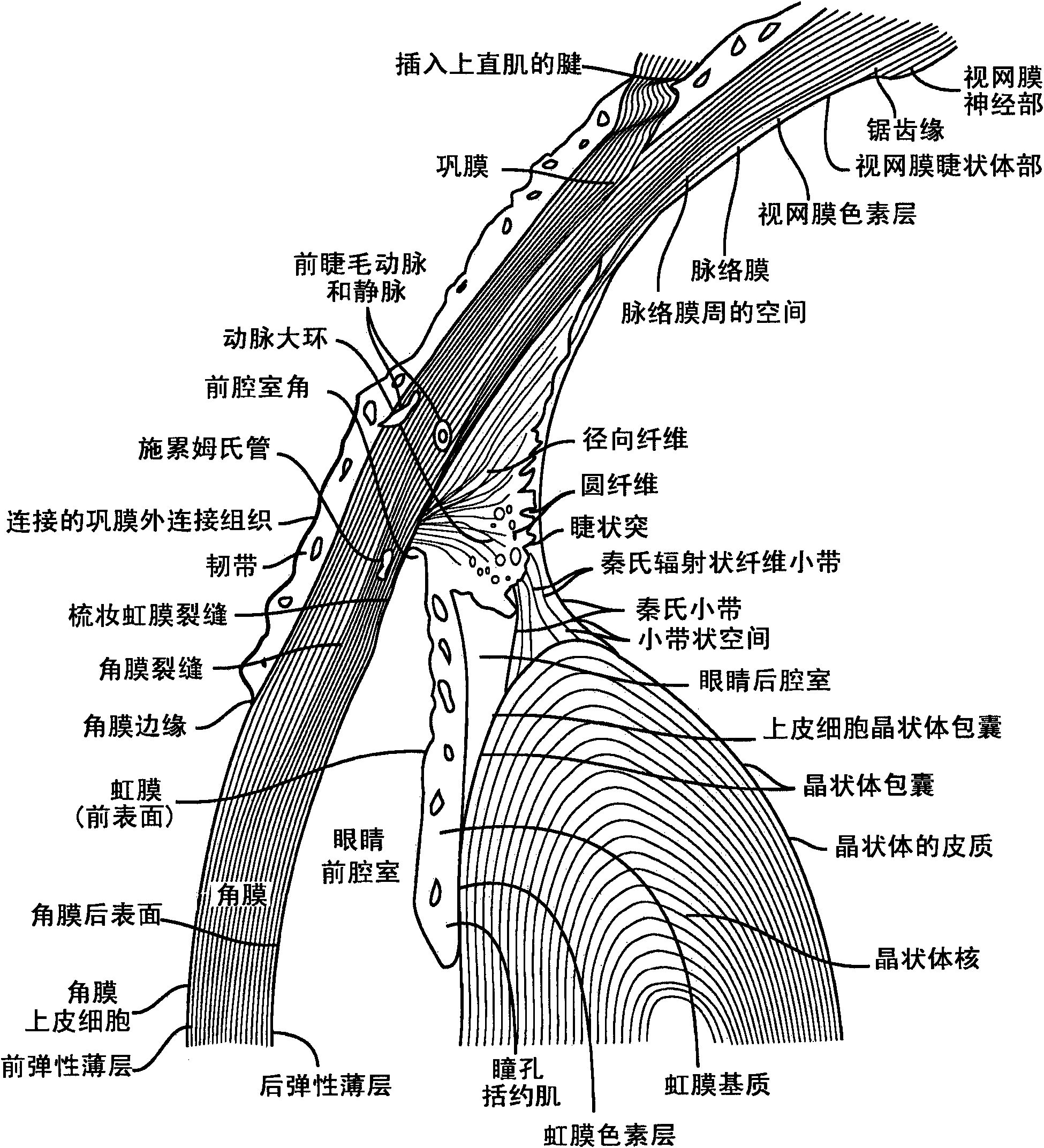
[0037] Embodiments described herein will find application in procedures for reducing intraocular pressure, such as draperectomy and trabeculectomy. Although the filtering procedure of dradenectomy is specifically mentioned, there are still new variations to this surgery that may be beneficial according to the embodiments described herein. For example, the methods and devices described herein can be used in a variety of procedures, including creating holes under the conjunctiva, blisters, and channels communicating with the anterior chamber so that aqueous fluid can drain from the anterior chamber, thereby reducing IOP. Also the systems, methods and devices described herein can be used as an aid in many procedures such as retinal surgery and cataract surgery, implants can be positioned at many parts of the eye to directly measure IOP, for example, one of the following or Multiple sites: intracorneal, anterior compartment, anterior, posterior compartment, posterior, vitreous and...


PUM


Abstract

Methods and apparatus for measurement of IOP following glaucoma surgery comprise an implant device having a pressure sensitive capacitor and coil sized for placement along the tissue drainage path, to monitor the success of the surgery and measure IOP directly. The implantable sensor device may comprise a MEMS based capacitive pressure sensor and coil. A complaint material is disposed over the pressure sensitive capacitor and coil to conform with tissue to further decrease invasiveness and such that the implant can measure pressure from at least a first side and a second side when positioned along the drainage path. The implant can work well with trabeculectomies and trabeculotomies, and can be positioned on the sclera at a location corresponding to the bleb, such that the effectiveness of the surgery and medication can be determined postoperatively to detect pressure changes and elevations.

Description

[0001] Cross-references to related applications [0002] This application claims priority to U.S. Patent Application No. 61 / 243,847, filed September 18, 2009, and entitled "Implantable Ophthalmic MEMS Sensing Device and Method," and US Patent Application No. 61 / 335,562, entitled "Implantable Ophthalmic Micro-Electro-Mechanical System Sensing Devices and Methods of Eye Surgery," filed May 8, the entire contents of which are incorporated herein by reference. [0003] Statement Concerning Rights to Inventions Acquired Under Search and Development Provided by the Federation [0004] Not obtained [0005] References to "sequence listings", listings or computer program listings attached on CD-ROM were not obtained Background of the invention [0006] Humans like to watch. Eyes are complex organs that allow a person to see his or her surroundings. The eye consists of the cornea and lens, which form images on the retina of the eye. The retina of the eye senses the light image for...

Claims


Application Information

Patent Timeline
Patent Type & Authority Applications(China)
IPC IPC(8): A61B3/16
CPCA61B3/16A61B2562/0247A61F9/00781A61B5/0031A61B2562/028
Inventor V·G·翁D·A·L·李
Owner ORTHOMEMS
Why Eureka
Try Eureka