Implantable ophthalmic MEMS sensor devices and methods for eye surgery

Inactive Publication Date: 2012-09-06
ORTHOMEMS
View PDF0 Cites 28 Cited by
  • Summary
  • Abstract
  • Description
  • Claims
  • Application Information

AI Technical Summary

Benefits of technology

[0009]Embodiments of the present invention provide methods and apparatus for improved glaucoma surgery and improved measurements of IOP following the glaucoma surgery. The apparatus may comprise an implant device having a transducer coupled to transmission circuitry, such as a pressure sensitive capacitor and a coil, respectively, each sized such that the implant can be used as an adjunct to surgery. The implantable sensor device can be sized such that the implanted sensor can be positioned at a location along a surgically created drainage path, for example between the sclera and conjunctiva, so as to monitor the outcome of the surgery and decrease invasiveness of the implant. As the implant can be localized to scleral tissue or positioned between the sclera and conjunctiva, sensitivity of the eye to trauma can be decreased. The implant may comprise a MEMS based capacitive pressure sensor and coil, such that the implant can be sized to decrease invasiveness. Also, the implant may comprise a complaint material disposed over the pressure sensitive capacitor and coil

Problems solved by technology

Unfortunately, ocular diseases can compromise vision of the eye and may cause blindness in at least some instances.
Glaucoma is a major cause of blindness in the United States.
Glaucoma is usually associated with an increase in intraocular pressure (hereinafter “IOP”), that can result in damage to the retina of the eye.
In at least some instances patient compliance is less than ideal, as can miss patients scheduled appointments, such that the IOP is not measured in a timely manner and appropriate therapy and treatment may be delayed.
Although treatment can be effective in many instances, in at least some patients may continue to lose vision under physician directed care.
Although measurements with an external IOP sensor can be helpful, these devices that Measure pressure of the eye with an external sensor are somewhat indirect and can be inaccurate in at least some instances, such that the measured IOP may differ from the actual pressure inside the eye.
In at least some instances, such assumptions can lead to errors in the indirectly measured

Method used


Image

  • Implantable ophthalmic MEMS sensor devices and methods for eye surgery
  • Implantable ophthalmic MEMS sensor devices and methods for eye surgery
  • Implantable ophthalmic MEMS sensor devices and methods for eye surgery

Examples


Embodiment Construction

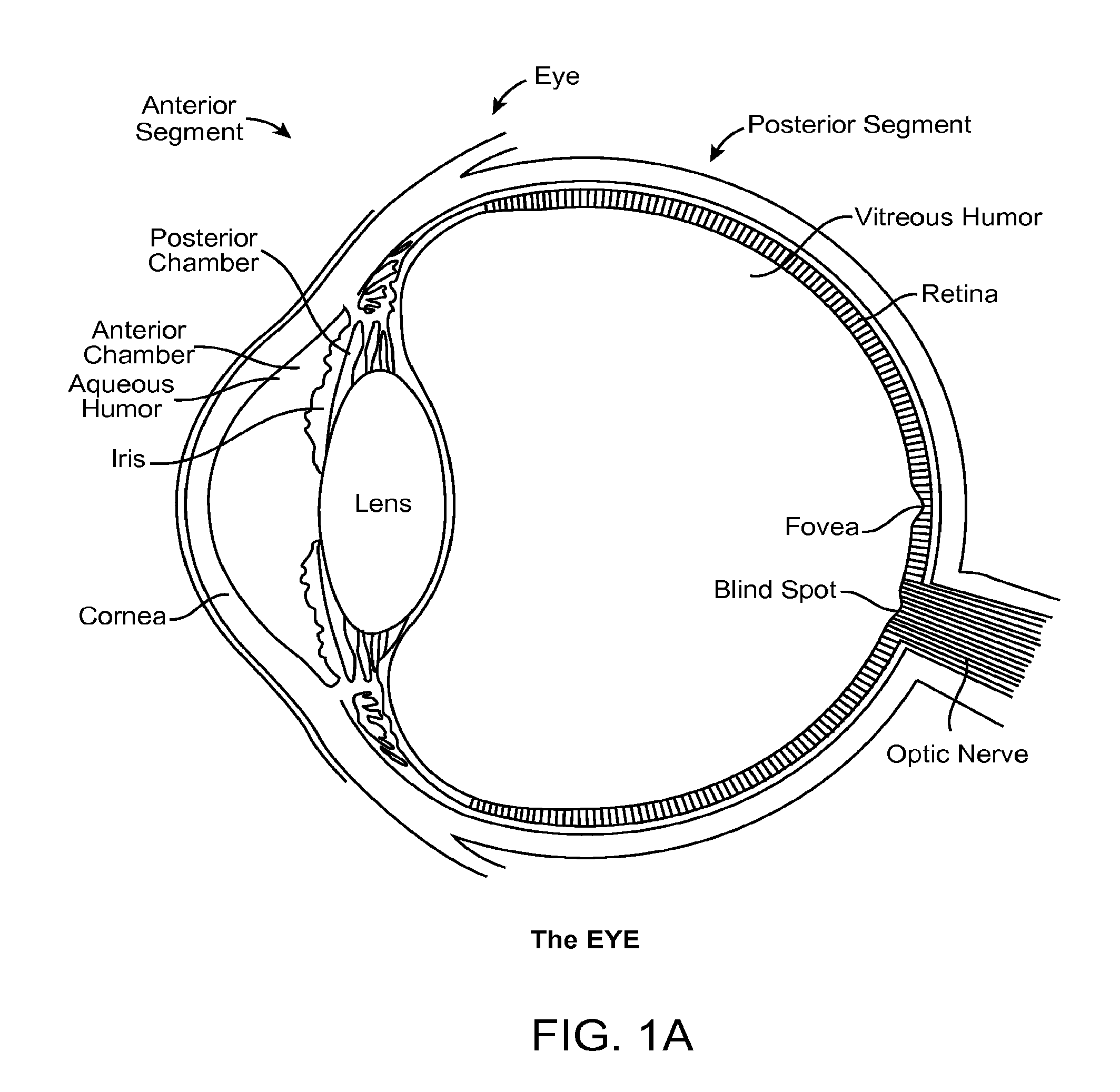
[0032]Embodiments described herein will have application to surgery to reduce intraocular pressure, for example with trabeculectomy and trabeculotomy. Although specific reference is trabeculectomy filtering surgery, there are new variations to this surgical procedure that can benefit in accordance with embodiments described herein. For example, the methods and apparatus described herein can be used with surgeries that involve forming a hole under the conjunctiva, a bleb, a scleral flap, and a channel communicating with the anterior chamber, such that aqueous fluid can drain from the anterior chamber, thereby lowering IOP. Also, the systems, methods and devices as described herein can be used as an adjunct with many surgeries such as retinal surgery and cataract surgery, and the implant can be positioned at many locations of the eye to directly measure IOP, for example one or more of intracorneal, anterior chamber, anterior segment, posterior chamber, posterior segment, vitreous and ...


PUM


Abstract

Methods and apparatus for measurement of IOP following glaucoma surgery comprise an implant device having a pressure sensitive capacitor and coil sized for placement along the tissue drainage path, to monitor the success of the surgery and measure IOP directly. The implantable sensor device may comprise a MEMS based capacitive pressure sensor and coil. A complaint material is disposed over the pressure sensitive capacitor and coil to conform with tissue to further decrease invasiveness and such that the implant can measure pressure from at least a first side and a second side when positioned along the drainage path. The implant can work well with trabeculectomies and trabeculotomies, and can be positioned on the sclera at a location corresponding to the bleb, such that the effectiveness of the surgery and medication can be determined postoperatively to detect pressure changes and elevations.

Description

CROSS-REFERENCES TO RELATED APPLICATIONS[0001]The present application is a continuation of PCT Application No. PCT / US / 2010 / 049527, filed on Sep. 20, 2010, which claims priority to the following patent applications: U.S. Pat. App. Ser. No. 61 / 243,843 filed on Sep. 18, 2009, entitled “Implantable Ophthalmic MEMs Sensor Devices and Methods”, and U.S. Pat. App. Ser. No. 61 / 335,572, filed on 8 Jan. 2010, entitled, “Implantable Ophthalmic MEMs Sensor Devices and Methods for Eye Surgery”, the full disclosures of which are incorporated herein by reference.BACKGROUND OF THE INVENTION[0002]People like to see. The eye is a complex organ that allows a person to see his or her surroundings. The eye includes a cornea and crystalline lens that form an image on the retina of the eye. The retina of the eye senses the light image formed thereon and transmits neural signals via the optic nerve to the occipital cortex of the brain, such that the person can see and perceive his or her surroundings. Unfo...

Claims


Application Information

Patent Timeline
IPC IPC(8): A61B3/16
CPCA61B3/16A61B5/0031A61F9/00781A61B2562/028A61B2562/0247
Inventor WONG, VERNON G.LEE, DOUGLAS A.
Owner ORTHOMEMS
Why Eureka
Try Eureka