Brain, carotid artery and aorta three-in-one scanning method and scanning system

A three-in-one, scanning method technology, applied in the field of biomedicine, can solve the problem of limited scanning time in the application of MRI technology, and achieve the effect of shortening the scanning time and reducing the scanning time

Active Publication Date: 2012-10-17
SHANGHAI UNITED IMAGING HEALTHCARE
View PDF5 Cites 9 Cited by
  • Summary
  • Abstract
  • Description
  • Claims
  • Application Information

AI Technical Summary

Problems solved by technology

[0003] The traditional method is to use MRI (Magnetic Resonance Imaging, Magnetic Resonance Imaging) to examine the brain, internal carotid artery, and aorta, which take about half an hour respectively. Putting them together, it takes 1.5 hours to 2 hours to scan, so Long scanning time limits the clinical application of MRI technology

Method used


Image

  • Brain, carotid artery and aorta three-in-one scanning method and scanning system
  • Brain, carotid artery and aorta three-in-one scanning method and scanning system
  • Brain, carotid artery and aorta three-in-one scanning method and scanning system

Examples


Embodiment Construction

[0033] The technical solution will be described in detail below in conjunction with specific embodiments and accompanying drawings.

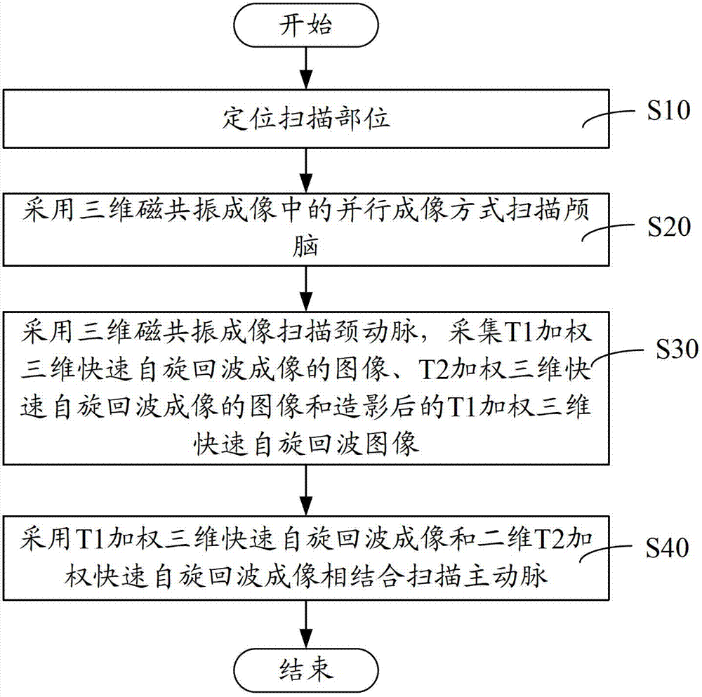
[0034] Such as figure 1 Shown, in one embodiment, a kind of scan method of brain, carotid artery and aorta Trinity, comprises the following steps:

[0035] Step S10, locating the scanning site.

[0036] Specifically, the head and neck radio frequency coil and heart coil are put on the patient, and the localizer is used for positioning scanning to obtain the localization scanning position, so that subsequent positioning scans do not need to be repeated, saving positioning time.

[0037] Step S20, scanning the cranium using parallel imaging in 3D magnetic resonance imaging.

[0038] Specifically, the parallel imaging method can be one of T1-weighted three-dimensional fast spin-echo imaging (T1-SPACE), T2-weighted three-dimensional fast spin-echo imaging (T2-SPACE) and three-dimensional magnetization preparative gradient echo sequence (MPRAGE) im...


PUM


Abstract

The invention relates to a brain, carotid artery and aorta three-in-one scanning method, comprising the following steps of: positioning a scanning part; scanning a brain by utilizing parallel imaging of three-dimensional magnetic resonance imaging; scanning a carotid artery by utilizing the three-dimensional magnetic resonance imaging and collecting a T1 weighted three-dimensional rapid spin echo imaged image, a T2 weighted three-dimensional rapid spin echo imaged image and a radiographed T1 weighted three-dimensional rapid spin echo image; and scanning a carotid artery by utilizing T1 weighted three-dimensional rapid spin echo imaging and T2 weighted three-dimensional rapid spin echo imaging which are combined. The brain, carotid artery and aorta three-in-one scanning method and system disclosed by the invention firstly adopt the steps of firstly positioning the scanning part and directly scanning the brain, the carotid artery and the aorta; the positioning and the scanning do not need to be repeated so that the scanning time is shortened; the brain is scanned by using a rapid imaging method by parallel imaging so that the scanning time is reduced; and three-dimensional T1 weighted imaging and two-dimensional T2 weighted imaging are combined to scan the carotid artery so that the scanning time is further shortened.

Description

【Technical field】 [0001] The invention relates to the field of biomedicine, in particular to a trinity scanning method and scanning system of cranium, carotid artery and aorta. 【Background technique】 [0002] Clinical studies have found that understanding the composition of plaques in the brain, carotid artery, and aorta is helpful for the diagnosis of cerebrovascular diseases. In order to understand the condition of the plaque inside the three, it is necessary to perform imaging examination on them respectively. [0003] The traditional method is to use MRI (Magnetic Resonance Imaging, Magnetic Resonance Imaging) to examine the brain, internal carotid artery, and aorta, which take about half an hour respectively. Putting them together, it takes 1.5 hours to 2 hours to scan, so Long scanning time limits the clinical application of MRI technology. 【Content of invention】 [0004] Based on this, it is necessary to provide a trinity scanning method of the cranium, carotid ar...

Claims


Application Information

Patent Timeline
Patent Type & Authority Applications(China)
IPC IPC(8): A61B5/055G06T7/00
Inventor 钟耀祖张磊张娜刘新郑海荣
Owner SHANGHAI UNITED IMAGING HEALTHCARE
Why Eureka
Try Eureka