Image-display device and capsule-type endoscope system

An image display device and capsule endoscope technology, which can be applied in the fields of endoscopy, image enhancement, image analysis, etc., can solve problems such as heavy burden on the interpreter, and achieve the effect of efficient interpretation

Inactive Publication Date: 2012-10-24
OLYMPUS CORP
View PDF11 Cites 9 Cited by
  • Summary
  • Abstract
  • Description
  • Claims
  • Application Information

AI Technical Summary

Problems solved by technology

[0003] The number of in-vivo images taken in one examination is as high as about 60,000 (about 8 hours), so the burden on the interpreter is very heavy

Method used


Image

  • Image-display device and capsule-type endoscope system
  • Image-display device and capsule-type endoscope system
  • Image-display device and capsule-type endoscope system

Examples


Embodiment approach 1

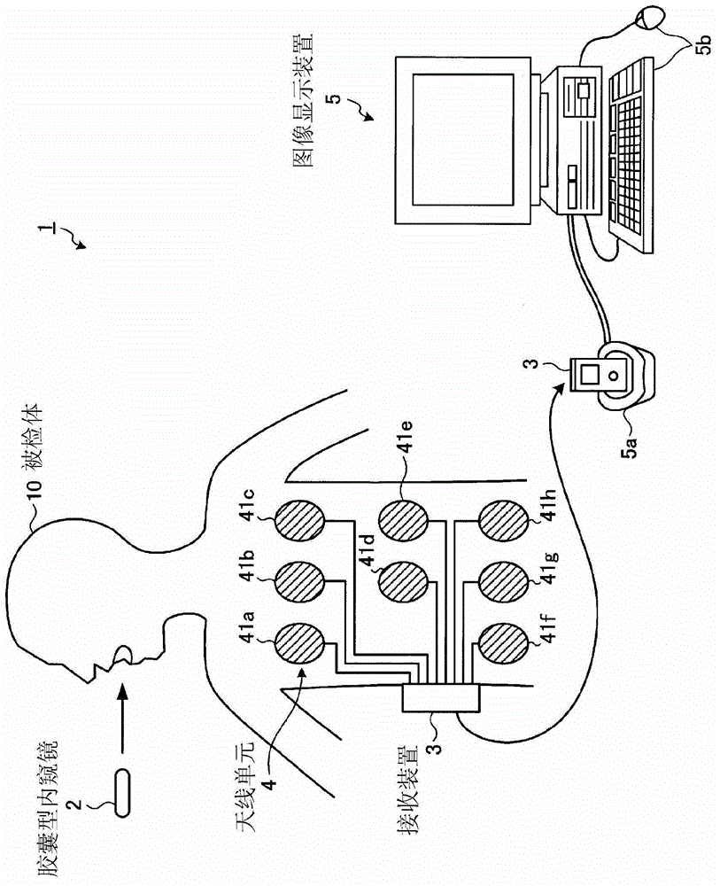
[0058] figure 1 It is a schematic diagram showing a schematic configuration of the capsule endoscope system according to Embodiment 1 of the present invention. The capsule endoscope system 1 includes: a capsule endoscope 2 that is introduced into the body of the subject 10 to take pictures and wirelessly transmits image data of in-vivo images to a receiving device 3; In-vivo image data wirelessly transmitted by the capsule endoscope 2 ; and an image display device 5 that displays the in-vivo image on a screen based on the in-vivo image data received by the receiving device 3 .

[0059] After the capsule endoscope 2 is swallowed from the mouth of the subject 10, it moves inside the organ of the subject 10 according to the peristaltic movement of the organ, etc. seconds) to sequentially image the inside of the subject 10 to obtain imaging signals, perform predetermined signal processing, and generate in-vivo image data. In addition, every time the capsule endoscope 2 takes an...

Deformed example 1-1

[0113] Next, refer to Figure 10 A first modified example of the image display device according to Embodiment 1 will be described. In the first embodiment described above, the user is made to arbitrarily select the type of image processing to be executed, but the user may be made to select the precision of various image processing. Figure 10 It shows the process selection screen displayed in this modification. In addition, in this modified example 1-1, sampling density information corresponding to the accuracy of various image processing is stored in advance in the storage unit 55 as a parameter.

[0114] if in Figure 10 When a predetermined operation signal (for example, aligning the cursor 131 with one of the icons 101 to 104 and right-clicking the mouse) is input on the processing selection screen 130 shown, the display control unit 59 causes the display unit 60 to display a selection image. A precision selection window 132 for processing precision (for example, three ...

Deformed example 1-2

[0118] Next, refer to Figure 11 and Figure 12 A second modified example of the image display device according to Embodiment 1 will be described. Figure 11 It is a block diagram showing the configuration of an image display device according to Modification 1-2. compared to Figure 4 , this image display device 5 - 2 further includes a trajectory calculation unit 61 in a subsequent stage of the position estimation unit 54 . Other structures are the same as those of the image display device 5 .

[0119] The trajectory calculation unit 61 executes trajectory calculation processing of the capsule endoscope 2 based on estimated position information obtained by the position estimation unit 54 . Specifically, the trajectory calculation unit 61 extracts two temporally adjacent points from the estimated plurality of positions of the capsule endoscope 2, and when the distance between the two points is equal to or less than a predetermined value, the two points are The points are ...


PUM


Abstract

Provided is an image-display device and the like that can start reading an image soon after obtaining image data captured by a capsule-type endoscope. The image-display device comprises: an I / F unit (51) for receiving information input for the image-display device; a storage unit (55) for storing in-vivo image data and information related to the position of the capsule-type endoscope in a subject's body; an image processor (53) for processing images for the in-vivo image data; a position estimator (54) for estimating the position of the capsule-type endoscope when in-vivo images are captured on the basis of the position-related information; a process-setting unit (56) for setting the content of image processing and the position estimation level of position estimation processing on the basis of information received from an input receiver; a processing-time forecasting unit (57) for forecasting the processing time required for processing by the image processor and the position estimator on the basis of the content of image processing and the position estimation level set by the process-setting unit; and a display unit (60) for displaying the processing time forecasted by the processing-time forecasting unit.

Description

technical field [0001] The present invention relates to an image display device and a capsule endoscope system for displaying in-vivo images obtained by a capsule endoscope introduced into a subject. Background technique [0002] Conventionally, when a capsule endoscope is introduced into the subject to take images of the subject, the following operations are performed: the internal images obtained by the capsule endoscope are viewed through a list of virtual moving images or still images. The image group is observed, and the image whose observation result is abnormal is selected. This operation is called interpretation. [0003] Since the group of in-vivo images taken in one examination is as high as about 60,000 (about 8 hours), the burden on the interpreter is very heavy. Therefore, as an auxiliary function for interpretation, for example, a function has been proposed (for example, refer to Patent Documents 1 to 3): to detect abnormal parts such as hemorrhage and tumor ...

Claims


Application Information

Patent Timeline
Patent Type & Authority Applications(China)
IPC IPC(8): A61B1/00A61B1/04G16H30/20G16H30/40
CPCG06T2207/10016G06F19/00G06T7/0012A61B5/061A61B1/041G06T2207/30028G06F19/321A61B1/00016G06F17/18G06T2207/30096G06T7/00G06T2207/10068G06F19/3406G06T2207/10024G16H40/63G16H30/40G16H30/20
Inventor 小林聪美高杉启
Owner OLYMPUS CORP
Why Eureka
Try Eureka