Nuclear medical imaging method and device

An imaging method and an imaging device technology, which are applied in the field of nuclear medical imaging and nuclear medical imaging devices, and can solve the problem that PET images are not optimal

Active Publication Date: 2012-10-31
TOSHIBA MEDICAL SYST CORP
View PDF5 Cites 26 Cited by
  • Summary
  • Abstract
  • Description
  • Claims
  • Application Information

AI Technical Summary

Problems solved by technology

Therefore, the resulting PET image may not be optimal from a logical point of view

Method used


Image

  • Nuclear medical imaging method and device
  • Nuclear medical imaging method and device
  • Nuclear medical imaging method and device

Examples


Embodiment Construction

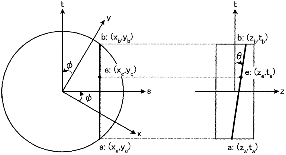
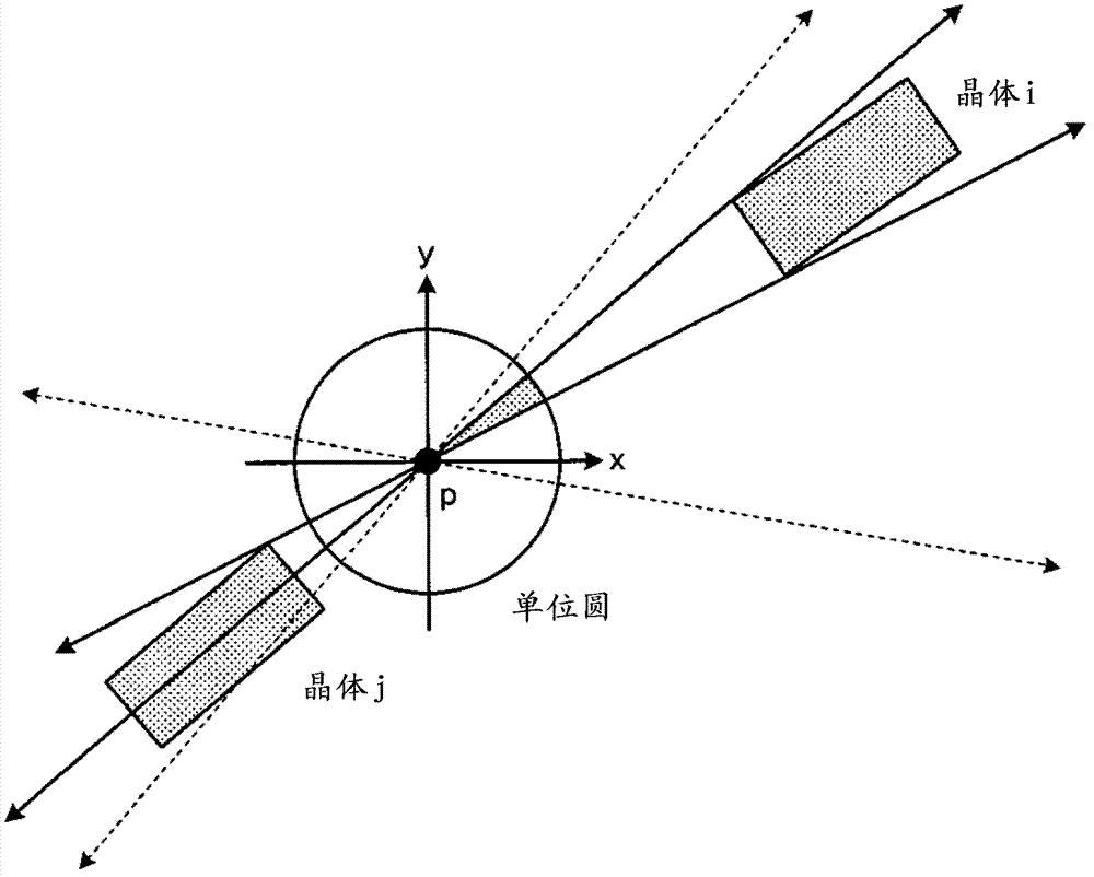
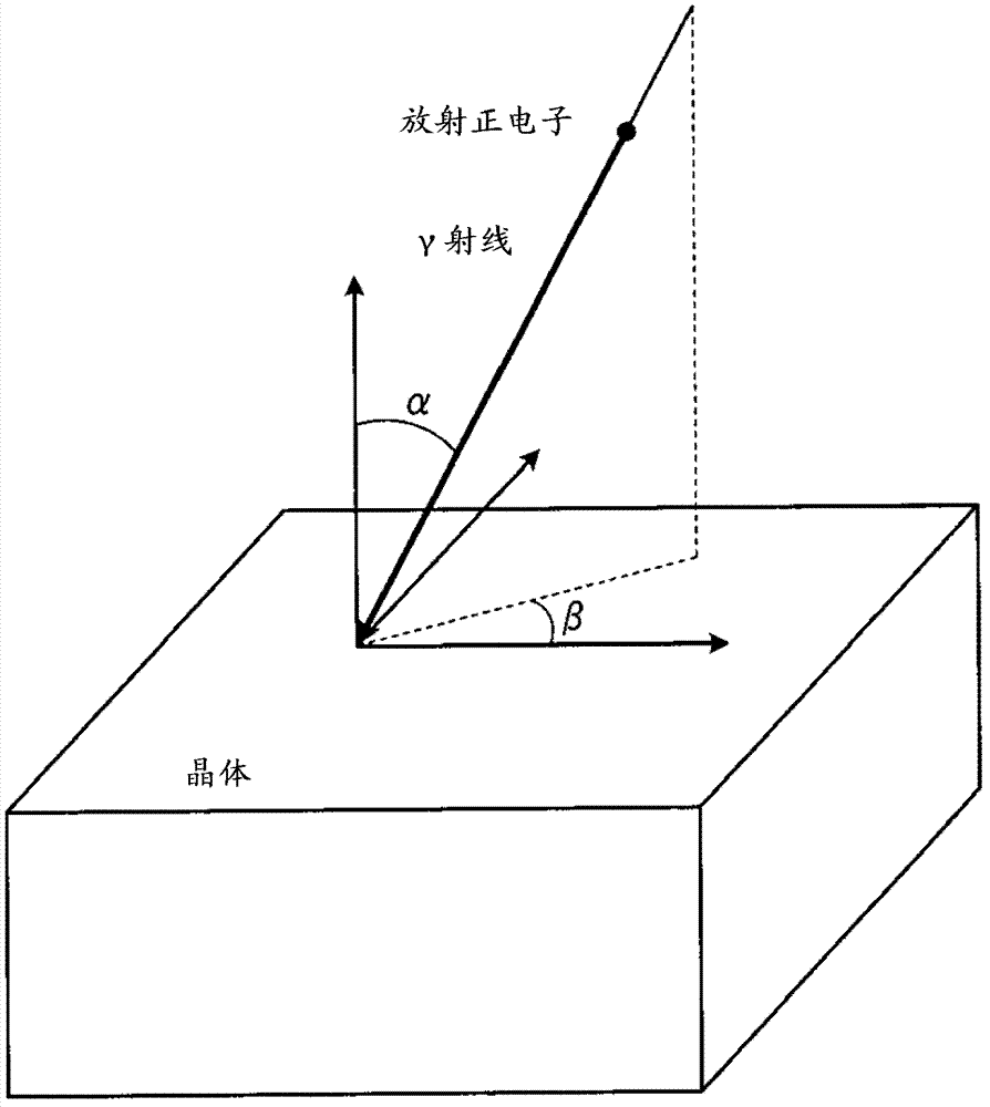
[0034] In one aspect of the embodiment, the nuclear medicine imaging method includes: (1) A first determination step of determining the LOR specified by the positions of a pair of detector crystals of a detector included in a PET imaging apparatus as a nuclear medicine imaging apparatus , (2) the definition step, which defines the array of radiation points corresponding to the determined LOR, (3) the second determination step, for each point in the array of radiation points corresponding to the above LOR, it is determined that the LOR will be defined The solid angle formed by the surface of the above-mentioned pair of detector crystals as the bottom surface, (4) the generating step, averaging the determined solid angles to generate an average solid angle, (5) the third determining step, the determination depends on the gamma ray To the DOI coefficient of the "position coefficient related to the position in the depth direction of interaction" transmitted through the pair of detec...


PUM


Abstract

The invention provides a nuclear medical imaging method and device for obtaining optimal images. The nuclear medical imaging method comprises a first determining step of determining a line of response set by the positions of a pair detector crystals, a defining step of defining an array of radiation points corresponding to the line of response, a second determining step of determining a solid angle with the bottom surface being surfaces of the pair of detector crystals defining the line of response for each point in the array of the radiation points corresponding to the line of response, a generating step of averaging the solid angle for generating an average solid angle, a third determining step of determining a position factor related to depth-of-interaction, and a calculation step of multiplying the reciprocal of the average solid angle by the position factor for calculating a geometric corrective factor for the line of response.

Description

[0001] This application claims the benefits of the priority of the U.S. Patent Application No. 13 / 096,672 filed on April 28, 2011 and the Japanese Patent Application No. 2012-85541 filed on April 4, 2012, and the above-mentioned patent applications are cited in this application all content. Technical field [0002] The embodiment of the present invention relates to a nuclear medicine imaging method and a nuclear medicine imaging apparatus. Background technique [0003] Positron Emission Tomography (PET) is a branch of nuclear medicine that introduces positron emission-type radioactive drugs into the body of the subject. When radiopharmaceuticals decay, positrons are generated. Specifically, in a phenomenon known as a pair of positron annihilation events (Positron annihilation) by reacting multiple positrons with electrons, respectively, a pair of positrons is generated while moving in substantially opposite directions along the coincidence line. γ (gamma) photon. The pairs of g...

Claims


Application Information

Patent Timeline
Patent Type & Authority Applications(China)
IPC IPC(8): A61B6/03
Inventor 董云王文莉
Owner TOSHIBA MEDICAL SYST CORP
Why Eureka
Try Eureka