Method for determining myofibrillar fragmentation index (MFI) by microscopy

A technology of myofibrils and flakes, which is applied in the field of measurement of important indicators of meat tenderization, can solve problems that affect the measurement results and cannot truly reflect the degree of myofibril fragmentation, and achieve the effect of reducing accidental errors

Inactive Publication Date: 2012-12-26
西安中基药用植物工程有限公司
View PDF0 Cites 3 Cited by
  • Summary
  • Abstract
  • Description
  • Claims
  • Application Information

AI Technical Summary

Problems solved by technology

Since the size of myofibril flakes has a significant impact on the protein concentration determination results of myofibril suspensions, the absorbance value cannot truly reflect the degree of myofibril flakes and directly affects the measurement results.

Method used


Image

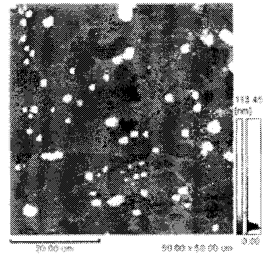
  • Method for determining myofibrillar fragmentation index (MFI) by microscopy
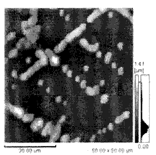
  • Method for determining myofibrillar fragmentation index (MFI) by microscopy
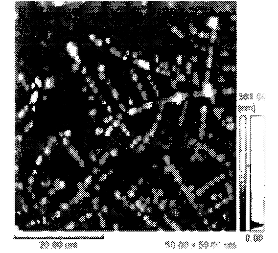
  • Method for determining myofibrillar fragmentation index (MFI) by microscopy

Examples


Embodiment Construction

[0013] According to the technical scheme of the present invention, the method for the microscopic measurement of myofibril fragmentation index of the present invention is carried out in the following manner:

[0014] First remove the visible fat and connective tissue in the meat sample, weigh about 5g, put it into a homogenizer, add 10mL of 2°C separation medium (the formula of the 2°C separation medium is: 100mmoL / L of KCL, 20mmoL / L of K 3 PO 4 , 0.1mmoL / L of EDTA, 1mmoL / L of CaCl 2 Mix to form a solution, then adjust to pH 7.0 with HCL), and homogenize at high speed and low temperature for 1 min. The homogenate was centrifuged at 3000g in a low-temperature centrifuge at 4°C for 15 minutes, and then the supernatant was slowly poured out to keep the precipitate. During the precipitation process, 10 mL of 2°C separation medium was added to the precipitate, and a suspension was made by stirring; Repeat the above centrifugation steps for the suspension, pour out the supernatant...


PUM


Abstract

The invention discloses a method for determining a myofibrillar fragmentation index (MFI) by microscopy. A meat sample without visual fat or connective tissue is put into a homogenizer, a separating medium with the temperature of 2 DEG C is added into the homogenizer for homogenating at a high speed and a low temperature, then supernatant liquor is slowly poured out, precipitate is retained, and the separating medium with the temperature of 2 DEG C is added into the precipitate again in the precipitation process to make a suspension with a stirring rod; and the steps are repeated for the prepared suspension, the supernatant liquor is poured out, the precipitate is still retained, the separating medium with the temperature of 2 DEG C is added into the precipitate and is mixed with the precipitate uniformly on a turbine mixer, the connective tissue is filtered out with a screen of 200 meshes, the separating medium with the temperature of 2 DEG C is added again to help myofibril to pass through sieve holes, the volume of the suspension is adjusted to be 10mL, the myofibril and the suspension are uniformly blended, 1 muL of myofibril suspension is sucked and dropped on a mica plate, and is naturally withered, an atomic power microscopic examination is directly carried out, and myofibrillar fragments are counted. The influence of the size of the myofibrillar fragment on a light absorption value in an absorption photometry of the existing method is avoided, and accidental errors are reduced.

Description

technical field [0001] The invention relates to the measurement of important indicators of meat tenderization, in particular to a method for microscopically measuring myofibril fragmentation index. This method improves the conventional determination method of myofibril fragmentation index, and can reflect the degree of myofibril fragmentation, that is, the tenderness of poultry meat, more directly and accurately. Background technique [0002] Tenderness is an important sensory characteristic of the eating quality of poultry and livestock meat, and it is also the main determinant of meat quality. At present, there are two main methods for measuring the tenderness of poultry meat: the shear force test method and the myofibril fragmentation index test method. Among them, the myofibril fragmentation index method has less accidental error than the shear force method, and is generally used at home and abroad. The essence of the myofibril fragmentation index method is to measure ...

Claims


Application Information

Patent Timeline
Patent Type & Authority Applications(China)
IPC IPC(8): G01Q60/24
Inventor 田成山
Owner 西安中基药用植物工程有限公司
Why Eureka
Try Eureka