Extending image information

An image and spectral information technology, applied in medical science, analysis using fluorescence emission, sensors, etc., can solve problems such as poor image contrast

CN102843953AInactive Publication Date: 2012-12-26KONINKLIJKE PHILIPS ELECTRONICS NV
10 Cites 0 Cited by

Patent Information

Authority / Receiving Office
CN · China
Patent Type
Applications(China)
Current Assignee / Owner
Publication Date
2012-12-26
Estimated Expiration
Not applicable · inactive patent

Smart Images

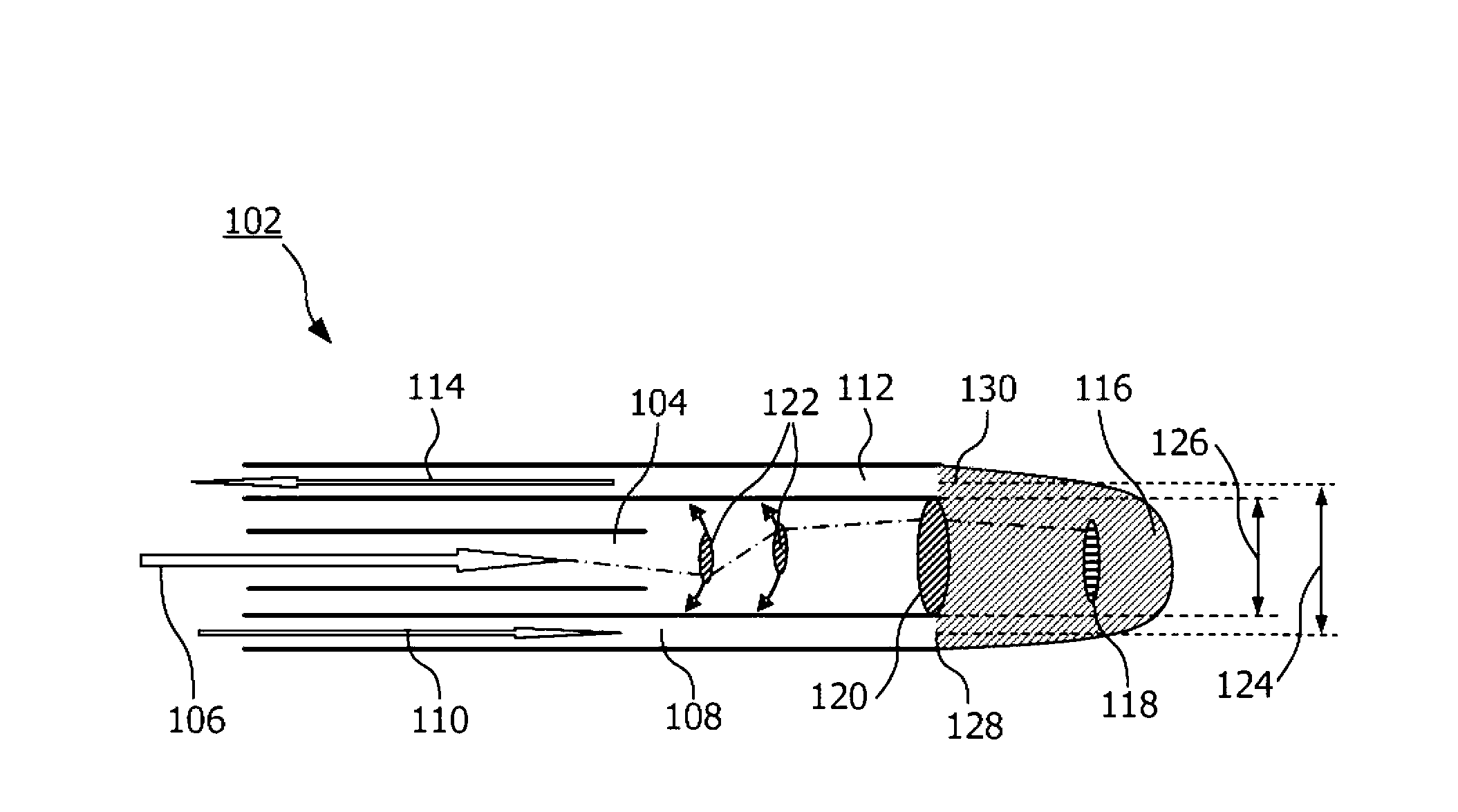
  • Figure 1
    Figure 1
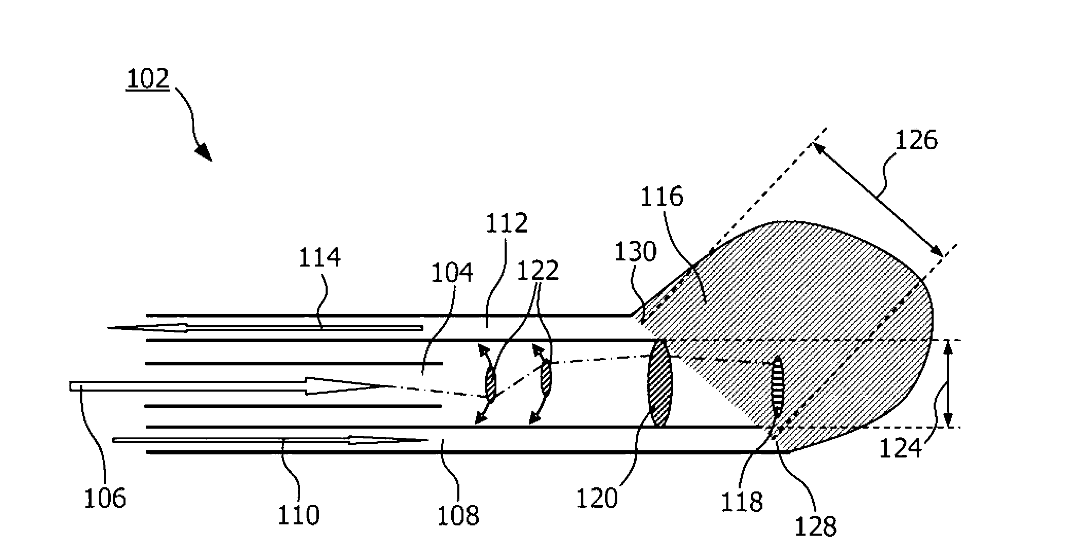
  • Figure 2
    Figure 2
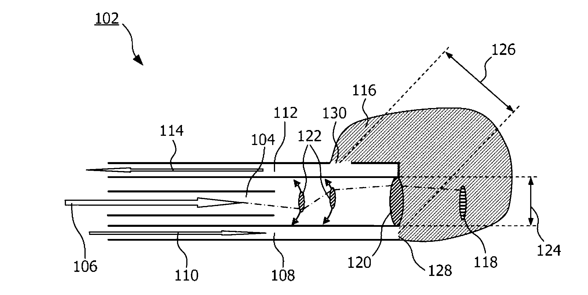
  • Figure 3
    Figure 3
Patent Text Reader

Abstract

The present invention relates to a system for extending microscopy information, where the microscopy information is image information from a first region 118 of an associated sample, the first region being imaged with an imaging system. The extension of the microscopy information originates from probing a larger second region 116 by photons which are emitted at an exit position 128 and collected at en entry position 130. The exit position and the entry position are spatially separated so that so that average spectral information of photons emitted from the exit position and collected at the entry position, is dependent on the second region 116 of the associated sample, the second region 116 being larger than the first region 118.
Need to check novelty before this filing date? Find Prior Art

Description

technical field

[0001] The present invention relates to the field of extended image information, in particular, the present invention relates to extended image information using averaged spectral information. Background technique

[0002] Interpretation of microscopic images of a sample may depend on parameters of the sample. For example, when imaging a portion of a sample, such as tissue or food, the sample may have a lipid-water ratio that is useful information for correct interpretation of the image. In oncology, the identification and grading of abnormal tissue from normal tissue is important. One way of obtaining this information is to take a biopsy and send this to the pathology department for diagnosis of the tissue. The problem here is that most of these biopsies are performed blindly, with no feedback on what tissue is in front of the biopsy device. Therefore, there is a risk of taking a biopsy in the wrong place.

[0003] Furthermore, sending a sample of tissue...

Examples

Embodiment Construction

[0075] figure 1 An embodiment of an interventional device 102 according to an embodiment of the invention is shown. The figure shows part of an interventional device, which may be part of a scanning fiber imaging system extended with two fixed fibers. The interventional device comprises a first guide part 108 and a second guide part 112 for guiding light. The imaging system comprises an imaging guide 104 through which photons can travel along such as in the direction depicted with arrow 6 . The imaging system also includes multiple lenses, such as fixed lens 120 and movable lenses such as 122 . In some embodiments, a moving lens may be coupled to imaging guide 104 that may move. The imaging system is capable and arranged to image a region of interest (ROI), also referred to here as a first region 118 . The first and second guides and their respective distal ends, the output location 128 and the input location 130, are spatially arranged such that when a photon moving throu...