
Automatic focusing method for ultrasonic elastography

A technology of ultrasonic elastography and autofocus, which is applied in the directions of ultrasonic/sonic/infrasonic image/data processing, ultrasonic/sonic/infrasonic Permian technology, organ movement/change detection, etc. Slow and other problems, to achieve the effect of increasing speed, reducing the amount of calculation, and good real-time application

Inactive Publication Date: 2013-02-27
SOUTH CHINA UNIV OF TECH
View PDF6 Cites 16 Cited by
  • Summary
  • Abstract
  • Description
  • Claims
  • Application Information

AI Technical Summary

Problems solved by technology

[0003] At present, the image strain calculation for elastography is carried out for the entire image, and the data volume is large, so the processing speed is slow, and there are certain limitations in clinical application.

Method used


Image

  • Automatic focusing method for ultrasonic elastography
  • Automatic focusing method for ultrasonic elastography
  • Automatic focusing method for ultrasonic elastography

Examples


Embodiment 1

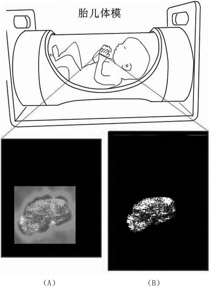
[0042] The implementation of the present invention requires ultrasonic equipment, which can directly run the program on the computer carried by the ultrasonic equipment, display the user graphical interface, and can adopt C++ language to compile various processing programs, so that the present invention can be better implemented. In this embodiment, an experiment is carried out. In the experiment, ultrasonic elastography of the region of interest is performed on the palm section of the fetal phantom. The operating environment of the Sonix RP system host of the ultrasonic equipment is: Pentium dual-core E2200CPU, the main frequency is 2.2GHz, and the memory is 1GB.

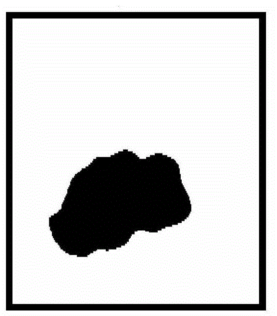
[0043] This example figure 1As shown, an autofocus method for ultrasound elastography, comprising the following steps:

[0044] (1) Ultrasound elastography was performed on the palm section of the fetal phantom, and the obtained elastic deformation image was normalized into a grayscale image, and then the grayscal...


PUM


Abstract

The invention discloses an automatic focusing method for ultrasonic elastography. The method comprises the following steps: normalizing and binarizing a plurality of obtained elastic displacement images; dividing the elastic displacement images into a plurality of image blocks; counting deformation amount of all image blocks; according to the deformation amount of each image block, dividing all the image blocks into two categories by using a cluster analysis: one is background and the other is an area of interest; and delimiting the final area of interest according to the classified image block information, defining the precomputed average displacement of the elastic images at a single line at the top of the area of the interest as the initial displacement of the area of interest, and carrying out elastic computation and imaging to only the area of interest during the following imaging process so as to realize automatic focusing. The automatic focusing method for ultrasonic elastography carries out elastic computation to ultrasonic radio frequency signals only in the area of interest, improves the imaging speed without influencing the imaging accuracy, can achieve the requirements of real-time application better and provides position information of underlying nidus for doctors automatically.

Description

technical field [0001] The invention relates to the technical field of computer-aided medical imaging, in particular to an automatic focusing method for ultrasonic elastography. Background technique [0002] Medical ultrasound diagnosis is an important part of modern medical imaging, and it has great reference value in the pathological diagnosis of many tissues of the human body (such as heart, liver, breast, superficial tissue, etc.). Changes in the elasticity or hardness of biological tissue are closely related to the abnormal pathological state of the tissue. When there is a hard object or mass inside the tissue, the elasticity or hardness of the tissue will change significantly. According to this principle, ultrasound-based elastography (Elastography) method can estimate the degree of deformation (or called strain, Strain) of the tissue under pressure or external dynamic excitation and vibration, and then can characterize the tissue more accurately flexibility. [0003...

Claims


Application Information

Patent Timeline
IPC IPC(8): A61B8/08
Inventor 黄庆华谢波
Owner SOUTH CHINA UNIV OF TECH
Learn More