Non-invasive standard configuration device suitable for ultrasound-computed tomography (CT)/magnetic resonance imaging (MRI)/position emission tomography (PET) and other fusion imaging
A non-invasive, -CT technology, applied in ultrasound/acoustic/infrasonic diagnosis, sonic diagnosis, infrasonic diagnosis, etc., can solve registration difficulties, technical difficulties and time-consuming problems of ultrasonic image fusion and registration
- Summary
- Abstract
- Description
- Claims
- Application Information
AI Technical Summary
Problems solved by technology
Method used
Image

Examples
Embodiment 1
[0064] Example 1 Rapid and Accurate Registration of Brain Surgery Navigation
specific example
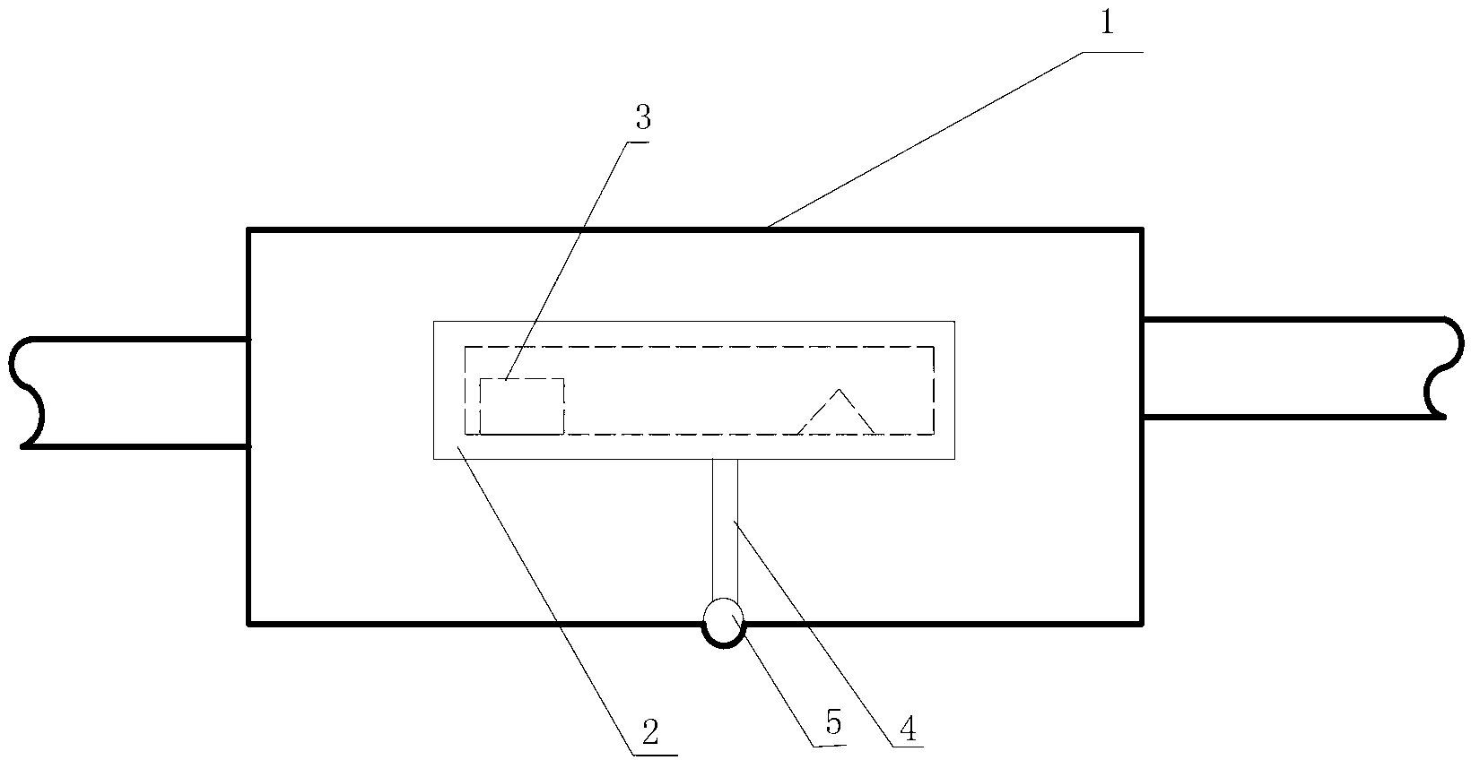
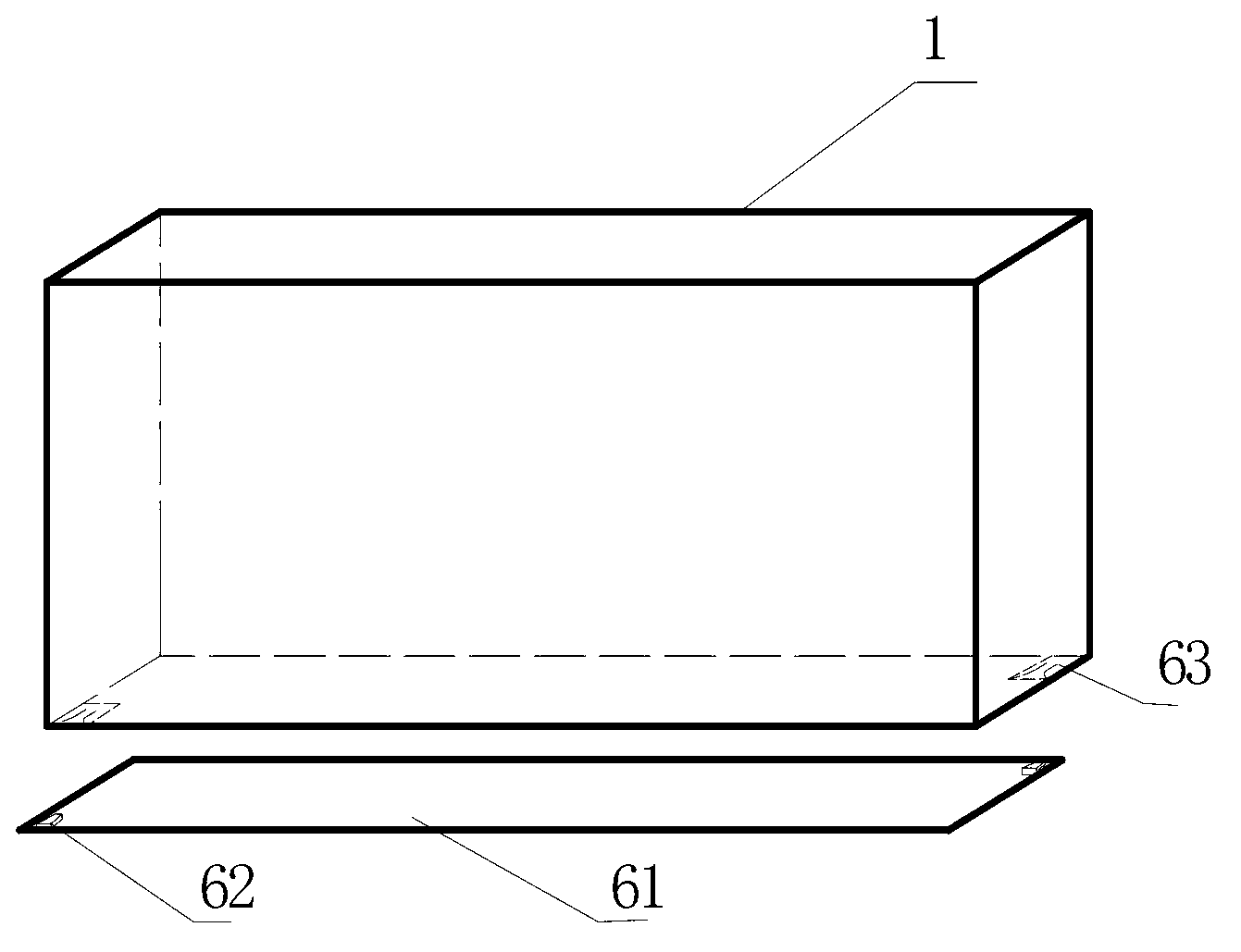
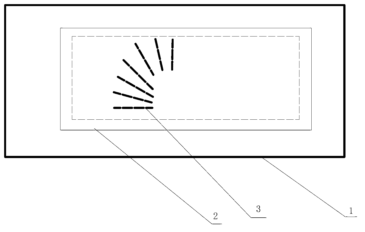
[0066] A 45-year-old male patient came to the doctor because of headache. MRI showed a left parasellar tumor with a diameter of 3 cm. Diagnosis: left parasellar meningioma. For accurate surgical positioning, ultrasound-MRI fusion imaging was performed. Before the MRI scan, the patient lies supine on the forehead and fixes the device of the present invention (the base of the device is a thin 3×4cm square with adhesive tape on one side, and a clip on each of the four corners of the non-adhesive side corresponding to the bayonet at the four corners of the lower end of the device body). MRI performs standard cross-sectional scans on the patient's head and the device of the present invention to obtain detailed intracranial images and detailed cross-sectional images of each geometric plane frame inside the device of the present invention. The next day, the patient's MRI DICOM image data was copied into the computer of the ultrasound fusion imaging system. In the electromagnetic fie...
Embodiment 2
[0071] Example 2 Guidance of radiofrequency ablation of liver tumors under the background of liver cirrhosis
[0072] 1. Specific examples
[0073]The patient is a 50-year-old male with 20 years of hepatitis B and liver cirrhosis. Both routine ultrasound and CT revealed a solid space-occupying tumor in the right liver with a diameter of 2cm. Diagnosis: primary liver cancer of the right liver. Ultrasound-guided radiofrequency ablation was proposed. For accurate surgical positioning, ultrasound-CT fusion imaging was performed. Before the CT scan, the patient was placed supine in the liver area to fix the device of the present invention (the base of the device is a thin 3×4 cm square with adhesive tape on one side, and a clip on each of the four corners of the non-adhesive side corresponding to the bayonet at the four corners of the lower end of the device body). CT performs standard cross-sectional scans on the abdomen of the patient and the device of the present invention to...
PUM
 Abstract
Description
Claims
Application Information