Organ geometry reconstruction method based on image volume element operation

A meta-operation and organ technology, applied in instrumentation, calculation, 3D modeling, etc., can solve problems such as low execution efficiency, ambiguous problem of model surface structure, analytical degree error, etc. Effect

Inactive Publication Date: 2013-04-24
HEFEI INSTITUTES OF PHYSICAL SCIENCE - CHINESE ACAD OF SCI
View PDF2 Cites 8 Cited by
  • Summary
  • Abstract
  • Description
  • Claims
  • Application Information

AI Technical Summary

Problems solved by technology

Based on high-precision human body image slices, commonly used 3D surface reconstruction algorithms (such as Marching Cubes) obtain the surface model structure by extracting the isosurface of the real object. On the one hand, due to the insufficiency of this surface extraction method, often It leads to the ambiguity of the surface structure of the model, resulting in many holes caused by the intersection of inside and outside or the error of the normal vector, which makes the model distorted and cannot truly express the internal geometric structure of the human body.
On the other hand, in the geometric reconstruction process, the analysis of the peripheral surface of the organ

Method used


Image

  • Organ geometry reconstruction method based on image volume element operation
  • Organ geometry reconstruction method based on image volume element operation
  • Organ geometry reconstruction method based on image volume element operation

Examples


Embodiment Construction

[0038] The present invention will be further described below in conjunction with the accompanying drawings and specific embodiments.

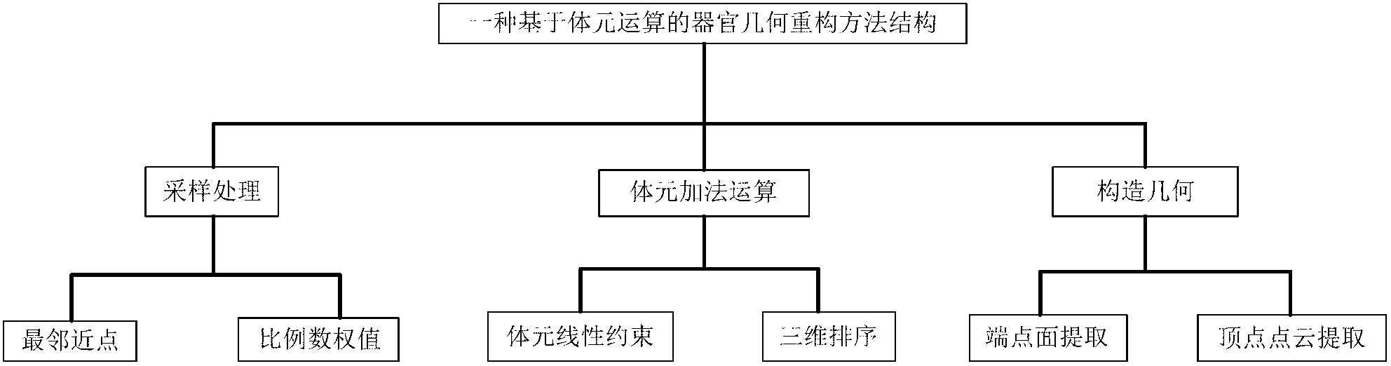
[0039] An organ geometric reconstruction method based on image voxel operation is a method for three-dimensional geometric reconstruction of human organs by generating a cuboid set of organs according to the voxel algorithm for the human body tomographic series slice data set after organ segmentation. In order to facilitate the understanding of this method, figure 1 A schematic diagram of the main function and method structure is provided, and the specific implementation steps are as follows:

[0040] Step 1.1 Obtain the position matrix of the specified organ voxel relative to the data set

[0041] The organ contours and color filling are performed on the tomographic series slice data, so that different organ voxels are given different RGB colors, and have a three-dimensional position relative to the data set, which is easy for computer identifi...


PUM


Abstract

The invention provides an organ geometry reconstruction method based on the image volume element operation. The method comprises the steps of acquiring a location matrix of a volume element relative data set of a specified organ, resampling the location matrix according to accuracy requirements, generating a cuboid coordinate matrix by means of the volume element additive operation, and reconstructing organ geometry according to the cuboid coordinate matrix. The human organ geometry reconstruction method is capable of quickly merging volume elements to the maximum level by means of a volume element additive operation algorithm, keeping the original shape of an organ, completely describing the detailed structure of the interior of the organ, reserving and controlling point clouds on the periphery surface of the organ by flexibly and efficiently combining with sampling and the volume element operation, and therefore Monte Carlo geometric description of an associated organ is enabled to be easy. The research result of the organ geometry reconstruction method based on the image volume element operation is applicable to efficient calculation of different Monte Carlo programs and assessment of human body radiation absorbed doses, and has wide application significance in fields like radiation safety or medical physics.

Description

technical field [0001] The invention relates to an organ geometry reconstruction method based on image voxel operation, through which a solid geometry model expressing the internal or external structure of a real object can be constructed efficiently. The calculation of dose absorption involving solid modeling and this geometric description has application value in the fields of radiation protection, medical radiotherapy planning and imaging science. Background technique [0002] Three-dimensional models are widely used in the fields of scientific experiment analysis and scientific experiment simulation. For example, in the field of nuclear radiation and protection, in order to simulate the absorbed dose of the human body under certain radiation conditions, it is often necessary to use computer technology to perform various processing on human body image data to establish an accurate human body geometric model, and combine it with Monte Carlo programs for simulation calculat...

Claims


Application Information

Patent Timeline
IPC IPC(8): G06T17/00
Inventor 赵凯吴宜灿黄群英龙鹏程程梦云
Owner HEFEI INSTITUTES OF PHYSICAL SCIENCE - CHINESE ACAD OF SCI
Why Eureka
Try Eureka