Demoscopy focus automatic segmentation method

An automatic segmentation and dermoscopy technology, applied in the field of medical image processing, can solve the problems of manual intervention and inaccurate segmentation, and achieve precise lesion analysis and diagnosis, accurate segmentation, and high efficiency

Inactive Publication Date: 2014-03-19
SHANXI UNIV
View PDF2 Cites 19 Cited by
  • Summary
  • Abstract
  • Description
  • Claims
  • Application Information

AI Technical Summary

Problems solved by technology

[0006] In view of the shortcomings of the existing dermoscopic lesion segmentation that requires manual intervention and the segmentation is not accurate

Method used


Image

  • Demoscopy focus automatic segmentation method
  • Demoscopy focus automatic segmentation method
  • Demoscopy focus automatic segmentation method

Examples


Embodiment Construction

[0027] The present invention will be described below in conjunction with the accompanying drawings and specific embodiments, but the present invention is not limited to this embodiment.

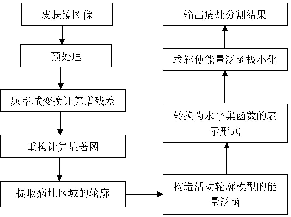
[0028] The automatic dermoscopy lesion segmentation method of the present invention utilizes the difference between the lesion area and its neighboring areas in the dermoscopic examination image to preliminarily divide the image, obtain the position, shape and contour information of the lesion area, and then construct the active contour based on these prior information The model is used for automatic segmentation of lesion regions, such as figure 1 As shown, the method includes the following steps:

[0029] For dermoscopy images with different types of lesions, the image is transformed in the frequency domain, and then the spectral residual is calculated to reconstruct the saliency map to obtain global saliency information. The lesion area and the background area in the image are initially di...


PUM


Abstract

The invention relates to a demoscopy focus automatic segmentation method in a medical image processing technology. The method includes the following steps: detection and extraction of a focus area; preliminary segmentation of a focus area and a background area in an image and extraction of the shape, position and contour of the focus area; segmentation of a mobile contour model; and construction of an initial curve of the mobile contour model according to the contour of the focus area and establishment of a corresponding energy functional. According to a level set method and a curve evolution theory, a curve capable of meeting minimum of the energy functional is obtained through partial-differential solution and the focus area is segmented automatically from the image. The method performs automatic detection and segmentation on the focus area according to the difference of the demoscopy focus area and the surrounding area; the method is accurate and high in efficiency in focus segmentation, great in practicality, dispensed with manual interruption and capable of assisting dermatology doctors to perform more accurate focus analysis and diagnosis.

Description

technical field [0001] The invention relates to the technical field of medical image processing, in particular to a method for automatically segmenting dermoscopic lesions. Background technique [0002] Dermoscopy, also known as skin surface light transmission microscope, is a non-invasive microscopic image analysis technique for observing the fine structure and pigments below the surface of living skin. It can observe the image structures and features invisible to the naked eye, such as the lower part of the epidermis, the papillary dermis, and the deep dermis. These features have a special and relatively clear correspondence with the changes in skin histopathology. Based on these correspondences, the dermoscopic diagnosis is determined. sensitivity and specificity. The latest research also shows that the specificity of dermoscopy in the diagnosis of malignant melanoma can reach 98%, even higher than clinical diagnosis. Therefore, dermoscopy is a fairly convenient, non-in...

Claims


Application Information

Patent Timeline
IPC IPC(8): G06T7/00G06T5/00A61B5/00
Inventor 王文剑白雪飞
Owner SHANXI UNIV
Why Eureka
Try Eureka