Multidimensional vein extracting method based on brain nuclear magnetic resonance image

A technology of nuclear magnetic resonance image, extraction method, applied in the field of medicine

Inactive Publication Date: 2014-05-14
CAPITAL UNIVERSITY OF MEDICAL SCIENCES
View PDF0 Cites 14 Cited by
  • Summary
  • Abstract
  • Description
  • Claims
  • Application Information

AI Technical Summary

Problems solved by technology

So far, no one has used the second-generation wavelet transform and Contourlet transform to extract the texture of AD brain MRI images.

Method used


Image

  • Multidimensional vein extracting method based on brain nuclear magnetic resonance image
  • Multidimensional vein extracting method based on brain nuclear magnetic resonance image
  • Multidimensional vein extracting method based on brain nuclear magnetic resonance image

Examples


Embodiment Construction

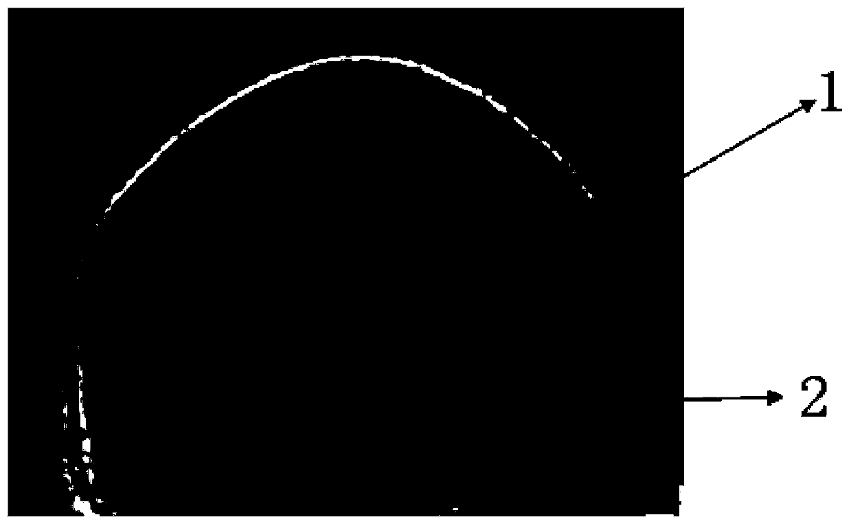
[0084] The following example is an introduction to using the method of the present invention to establish a model for predicting AD based on brain MRI images containing relevant ROIs. This is only a further description of the method of the present invention, but the examples do not limit the application scope of the present invention. In fact, this method can also be used to judge the properties of other types of medical images.

[0085] Image source: The brain MRI images of AD, MCI and normal elderly shared on the ADNI website are in .Nii format and read using MRIcro software;

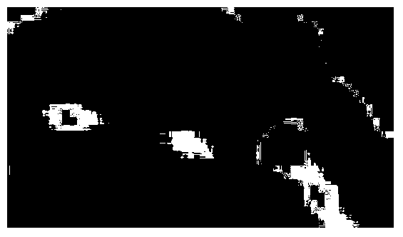
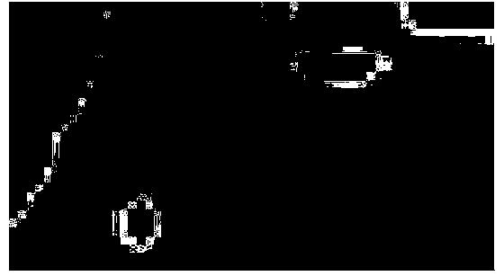
[0086] Methods: Using Matlab software programming, using region growth method to segment the ROIs in the above MRI image, and using Contourlet transform to extract the texture feature parameters of the relevant ROIs.

[0087] The following is an example of extracting texture feature parameters of ROIs from brain MRI images. The steps are as follows:

[0088] 1. Collect 250 original brain MRI images of 20 case...


PUM


Abstract

The invention discloses a multidimensional vein extracting method based on a brain nuclear magnetic resonance image. An area-of-interests in the brain nuclear magnetic resonance image is segmented through a region growing method. Vein characteristic parameters of the area-of-interests are extracted through a Curvelet conversion and Contourlet conversion method. The people comprise an Alzheimer patient group, a mild cognitive impairment patient group and normal old people group, the vein characteristic parameters of the area-of-interests comprise entropy, gray average, correlation, energy, the homogeneity degree, variance, the maximum probability, deficit distance, an inverse gap, clustering tendency, contrast, a sum mean value, a difference mean value, a sum entropy and a difference entropy. The area-of-interests comprises a entorhinal cortex are and a sea horse area.

Description

Technical field: [0001] The invention belongs to the field of medical technology, and specifically relates to a multi-dimensional texture extraction method based on brain magnetic resonance images (MRI). Background technique: [0002] In assisting the diagnosis of early Alzheimer's disease (AD), it is of great significance to identify the nature of ROIs (including entorhinal cortex and hippocampus) in MRI images. However, MRI imaging technology can only use hippocampal atrophy as one of the indicators to distinguish between patients and normal people. The interpretation of MRI images by doctors is easily affected by subjective individuals, lacks consistency, and it is difficult to accurately evaluate the severity of symptoms in dementia patients. [0003] 1. There are 5 types of existing image processing technologies: [0004] 1) Region-growing Method: [0005] This method uses the local spatial information of the image and can effectively overcome the shortcomings of the discontinuo...

Claims


Application Information

Patent Timeline
Patent Type & Authority Applications(China)
IPC IPC(8): G06K9/46G06K9/62G06V10/52G06V10/764
CPCG06V10/52G06V2201/03G06V10/764
Inventor 郭秀花高妮王晶晶陶丽新刘相佟
Owner CAPITAL UNIVERSITY OF MEDICAL SCIENCES
Why Eureka
Try Eureka