Blood vessel seed point selecting method and blood vessel extracting method in angiography

A seed point and blood vessel technology is applied in the field of medical tomographic image processing, which can solve the problems of manually selecting seed points and failing to achieve fully automatic blood vessel segmentation.

Active Publication Date: 2014-05-21
SHANGHAI UNITED IMAGING HEALTHCARE
View PDF5 Cites 24 Cited by
  • Summary
  • Abstract
  • Description
  • Claims
  • Application Information

AI Technical Summary

Problems solved by technology

It can be seen from the above that the common disadvantage of segmentation methods that are not based on standard vascular models is that they need to manually select seed points and cannot achieve fully automatic vascular segmentation.

Method used


Image

  • Blood vessel seed point selecting method and blood vessel extracting method in angiography
  • Blood vessel seed point selecting method and blood vessel extracting method in angiography
  • Blood vessel seed point selecting method and blood vessel extracting method in angiography

Examples


Embodiment Construction

[0038] The present invention will be further described below in conjunction with the accompanying drawings and embodiments.

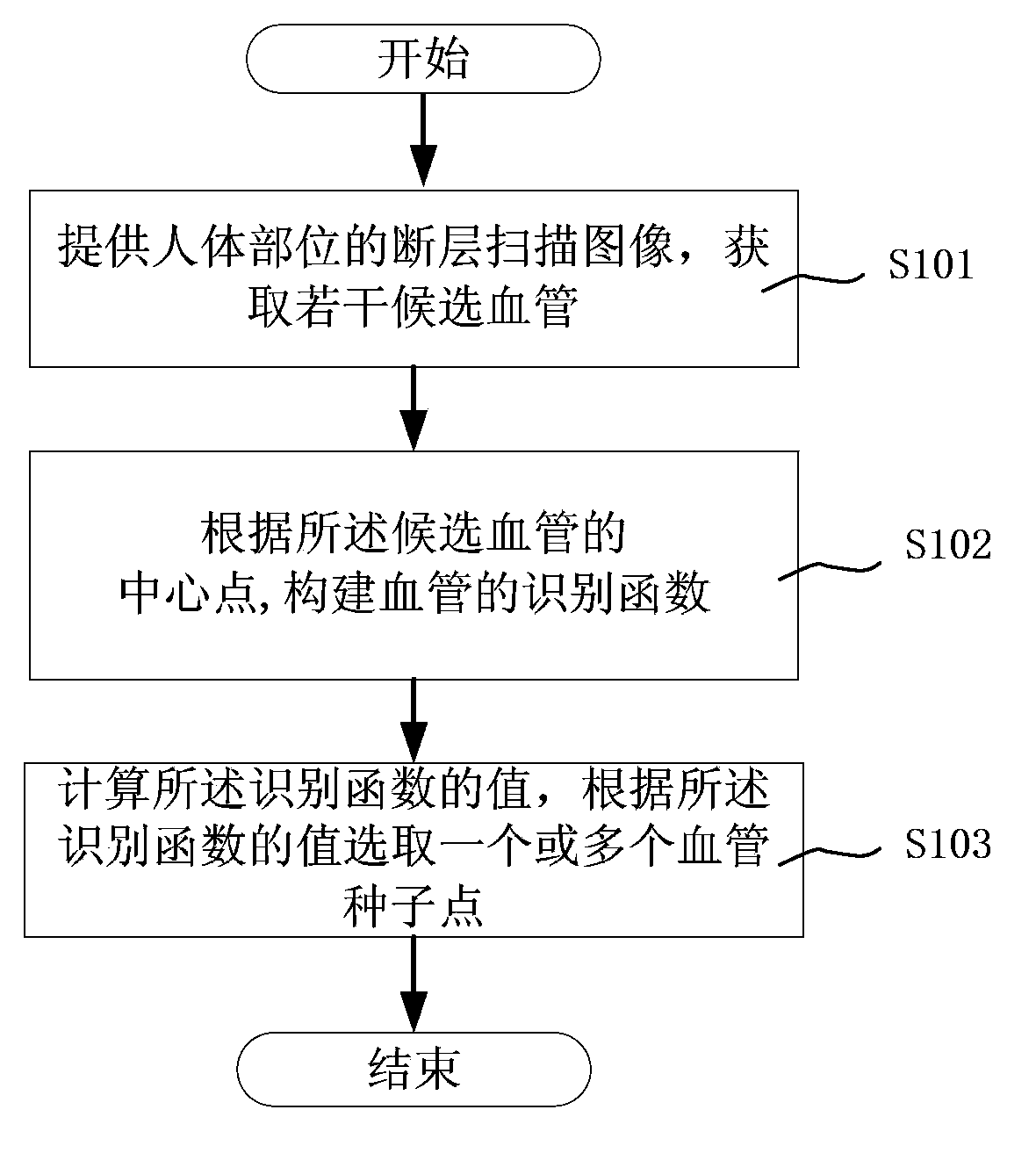
[0039] figure 1 It is a flowchart for selecting blood vessel seed points in angiography of the present invention.

[0040] See figure 1 , the blood vessel seed point selection method in the angiography provided by the present invention, comprises the following steps:

[0041] In step S101, a tomographic image of a human body is provided, and several candidate blood vessels are obtained. Before obtaining the plurality of candidate blood vessels and the center points of the candidate blood vessels, the method further includes: performing blood vessel enhancement on the tomographic image; and acquiring the plurality of candidate blood vessels and the center points of the candidate blood vessels according to the enhanced tomographic image. The blood vessels are enhanced by point regions based on Hessian matrix eigenvalues, or by Hough transform. Accordi...


PUM


Abstract

The invention discloses a blood vessel seed point selecting method and a blood vessel extracting method in angiography. The method comprises the following steps of providing a tomoscan image of a human body part and obtaining a plurality of candidate blood vessels; establishing an identification function of the candidate blood vessels according to the center points of the candidate blood vessels; calculating the value of the identification function and selecting one or more blood vessel seed points according to the value of the identification function. According to the blood vessel seed point selecting method in the angiography, the blood vessel seed points can be selected without the need of a standard model, and the method has the characteristics of high identification reliability and fast identification speed, and blood vessels can be fast extracted by finally combining a blood vessel fast dividing technology.

Description

technical field [0001] The invention relates to a processing method of a medical tomographic scanning image, in particular to a method for selecting a blood vessel seed point and a blood vessel extraction method in angiography. Background technique [0002] In Computed Tomography Angiography (CTA for short), it is necessary to separate blood vessels from other tissues for further observation and analysis of blood vessels. Existing techniques for vessel extraction in CTA are mainly divided into two categories: methods based on standard vessel models and methods not based on standard vessel models. [0003] US Patent Document 1: Jean-Michel Rouet, Franck Laffargue and Maxim Fradkin, Imageprocessing system for automatic segmentation of a 3-D tree-like tubular surface of an object, using 3-D deformable mesh models. US Patent, 2008, US 2008 / The variable mesh model method provided by 0094389A1 needs to provide a standard blood vessel model first, then calculate the centerline of...

Claims


Application Information

Patent Timeline
Patent Type & Authority Applications(China)
IPC IPC(8): G06F19/00G06T7/00
Inventor 王晓东李程毛玉妃
Owner SHANGHAI UNITED IMAGING HEALTHCARE
Why Eureka
Try Eureka