CT (Computed Tomography) value correcting method for cone-beam CT

A correction method and CT value technology, applied in the field of medical devices, can solve the problems of complex and time-consuming processes

Active Publication Date: 2014-08-06
NORTHEASTERN UNIV
View PDF7 Cites 35 Cited by
  • Summary
  • Abstract
  • Description
  • Claims
  • Application Information

AI Technical Summary

Problems solved by technology

Establish the corresponding relationship between the actual test CT value and its ideal CT value under various scanning conditions. In actual operation, multiple state curves need to be established, and the process is complicated and time-consuming.

Method used


Image

  • CT (Computed Tomography) value correcting method for cone-beam CT
  • CT (Computed Tomography) value correcting method for cone-beam CT
  • CT (Computed Tomography) value correcting method for cone-beam CT

Examples


Embodiment Construction

[0056] The specific implementation of the present invention will be described in detail below in conjunction with the drawings.

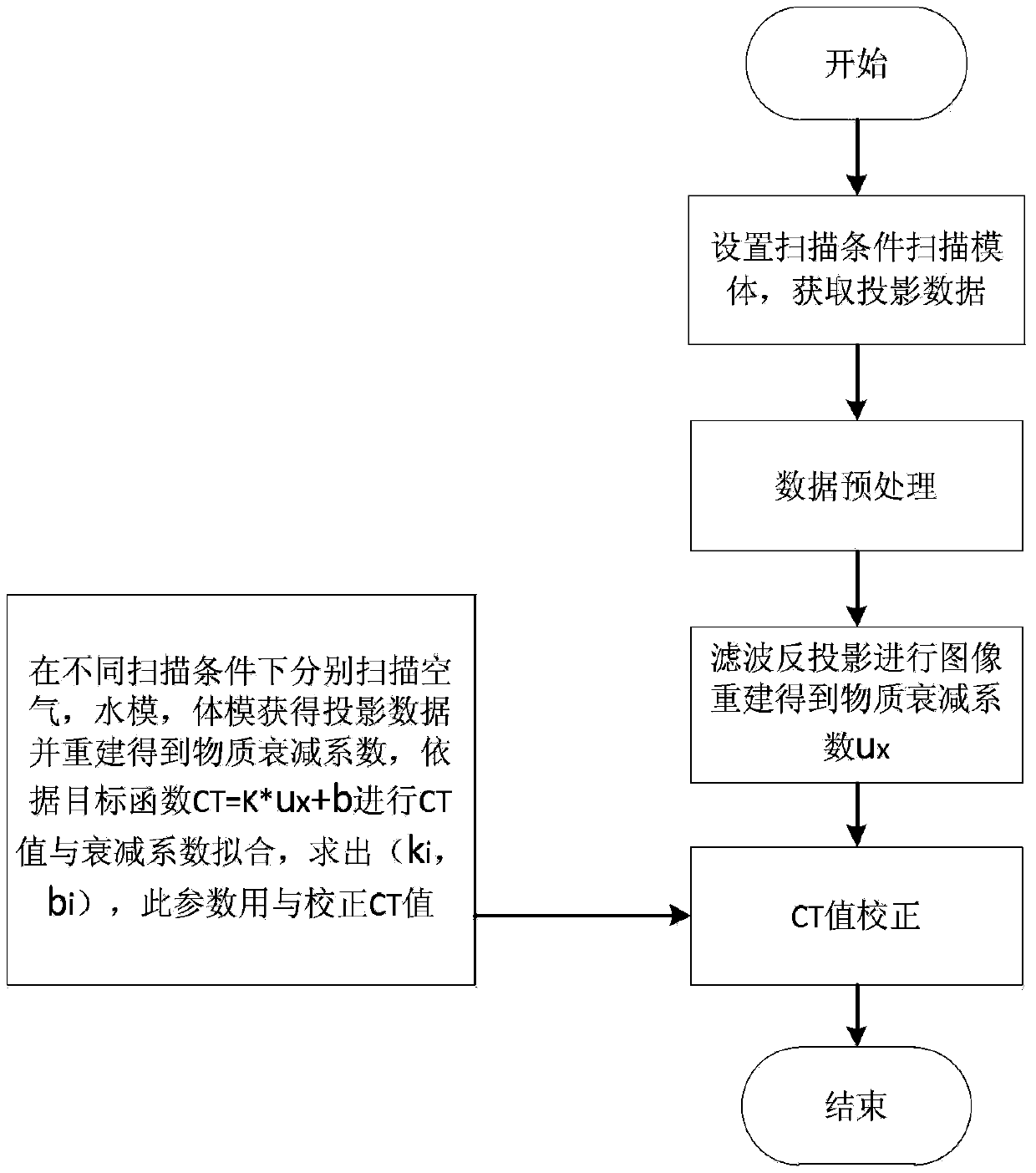
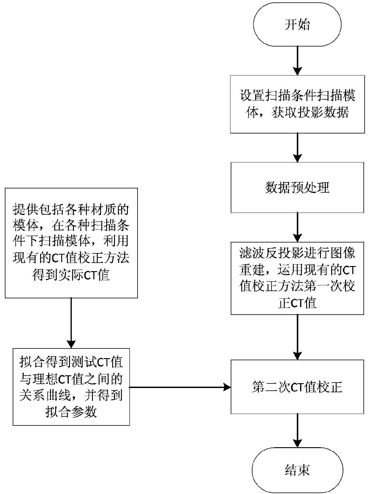
[0057] This embodiment is to apply the CT value image correction method for cone beam CT to oral cone beam CT, such as image 3 As shown, the CT value correction method for cone beam CT includes the following steps:

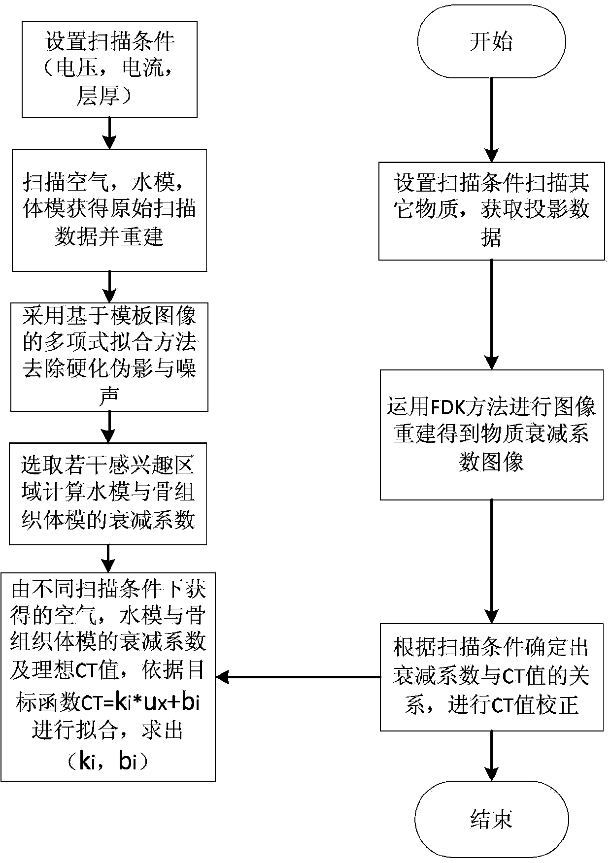
[0058] Step 1: Set cone beam CT scanning conditions, including current, voltage and slice thickness;

[0059] In the oral cone-beam CT used in this embodiment, there are two commonly used scanning conditions (group 1 and group 2), one group is 1 mA, 55 kV, and the thickness is 0.126 mm, and the second group is 2 mA, 60 kV. The layer thickness is 0.126 mm.

[0060] Step 2: Scan the air, water and bone tissue phantoms under different scanning conditions to obtain projection data of the air, water and bone tissue phantoms respectively;

[0061] The image reconstruction matrix is ​​512×512, the scanning field of view is 65mm, and the air, water and...


PUM


Abstract

The invention discloses a CT (Computed Tomography) value correcting method for cone-beam CT. The CT value correcting method comprises the following steps: scanning air, a water phantom and a bone tissue body phantom under different scanning conditions to respectively obtain projection data; reestablishing an image; removing noise and artifacts by adopting a polynomial fitting method based on a template image; selecting a plurality of interested regions and calculating a mean value of an attenuation coefficient and a standard deviation of the attenuation coefficient; obtaining the value range of the attenuation coefficient and taking the attenuation coefficient of maximum occurrence probability; fitting the attenuation coefficients of the air, the water phantom and the bone tissue body phantom under the different scanning conditions with corresponding ideal CT values to obtain respective fitting curves; performing CT scanning and image reestablishment on a scanned material; obtaining a CT value image according to a material attenuation coefficient image and a fitting curve and finishing the CT value correction. According to the CT value correcting method disclosed by the invention, image noise and artifact can be effectively reduced, the reestablished image of single material reaches the uniformity to the great degree, the precision of the attenuation coefficient of the material becomes higher and further high accuracy for the fitting of the curve of the CT value and the material attenuation coefficient is realized.

Description

Technical field [0001] The present invention relates to the technical field of medical devices, in particular to a CT value correction method for cone beam CT. Background technique [0002] CT examination is an indispensable examination method in formulating surgical plans and setting radiation doses. The correct CT diagnosis can only be concluded through the lesion information provided by high-quality CT photos. Therefore, the control of the quality of CT images has become a great concern The problem. In analyzing CT images, the changes in tissue density can be effectively understood by measuring CT values. If the CT value is not accurate enough, it is easy to cause misjudgment and cause the operation to fail. In radioactive RT therapy, the CT value is used as a reference for the radiation dose provided by the doctor. The larger the CT value, the greater the radiation dose required. Conversely, the smaller the CT value, the smaller the radiation dose required. If the CT value...

Claims


Application Information

Patent Timeline
Patent Type & Authority Applications(China)
IPC IPC(8): A61B6/03
Inventor 康雁孝大宇陈永丽田方俊徐礼胜
Owner NORTHEASTERN UNIV
Why Eureka
Try Eureka