Quantitative analysis method aiming at apoptotic cell morphology of fluorescence microscopic image

A technology of apoptotic cells and microscopic images, which is applied in the field of quantitative analysis of apoptotic cell morphology in fluorescent microscopic images, can solve the problems of indistinguishability and analysis, reduce human judgment differences, improve consistency, and segment speed fast effect

Inactive Publication Date: 2014-08-20
TIANJIN UNIV
View PDF5 Cites 13 Cited by
  • Summary
  • Abstract
  • Description
  • Claims
  • Application Information

AI Technical Summary

Problems solved by technology

[0006] In order to overcome the defect that the existing cell image analysis method cannot be combined with the staining results to distinguish and analyze normal cells and apoptotic cells, the present invention provides a two-dimensional morphological analysis method for apoptotic cells, which can not only Effectively segment the individual cells from the image, automatically calculate the two-dimensional morphological parameters of the cells, without the need for the user to manually outline the cells, greatly shortening the image analysis time, and can combine the staining results to classify and count apoptotic cells and normal cells , to provide a quantitative expression of the state of apoptosis using morphological characteristic parameters, providing modern means and more accurate data for the processing of cell images and the analysis of morphological differences between apoptotic cells and normal cells

Method used


Image

  • Quantitative analysis method aiming at apoptotic cell morphology of fluorescence microscopic image
  • Quantitative analysis method aiming at apoptotic cell morphology of fluorescence microscopic image
  • Quantitative analysis method aiming at apoptotic cell morphology of fluorescence microscopic image

Examples


Embodiment Construction

[0036] The technical solution adopted by the present invention to solve its technical problems is as follows:

[0037] The method for analyzing the two-dimensional morphology of apoptotic cells comprises the following steps:

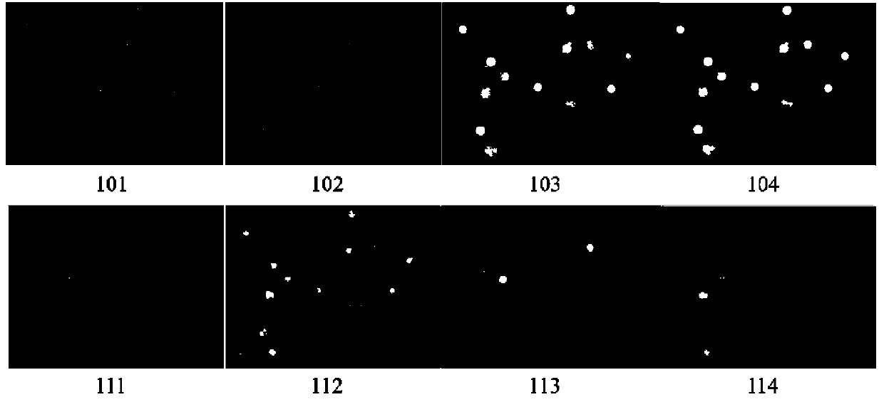
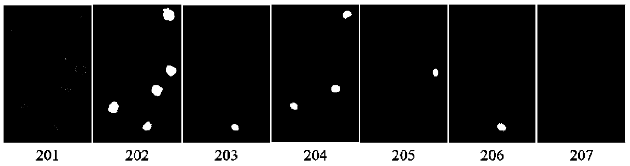
[0038]1) Acquire several pairs of fluorescent microscopic cell images to be processed. A pair of images includes a light microscope image and a three-stained fluorescent image for detecting cell apoptosis (Hoechst33342, bibenzyme nucleic acid dye, permeable The normal cell membrane enters the cell, and the staining result is blue; Annexin V, a phospholipid-binding protein, has a high affinity with phosphatidylserine, and it binds to the cell membrane of early apoptosis cells through the phosphatidylserine exposed on the outside of the cell, and the staining result is Green; PI, propidium iodide nucleic acid dye, cannot pass through the normal cell membrane, can only mark late apoptotic nuclei, and the staining result is red), the image contains normal ce...


PUM


Abstract

The invention relates to a quantitative analysis of an apoptotic cell morphology, which can effectively divide cell individuals from an image, automatically calculate cell two-dimensional morphology parameters and greatly shorten image analyzing time. The technical scheme adopted by the invention is a quantitative analysis method aiming at the apoptotic cell morphology of a fluorescence microscopic image. The quantitative analysis method comprises the following steps: (1) collecting a plurality of pairs of fluorescence microscopic cell images to be treated; (2) applying median filtering to a light microscope image in one pair of the images; carrying out gray stretching and carrying out image pre-treatment by using morphology opening operation; (3) automatically finding a threshold value by using an Otsu algorithm; (4) obtaining a divided binary image; (5) filling holes by using a morphology expansion and corrosion method, and removing small regions and incomplete targets on the boundary; (6) judging the state of a cell by the divided binary image according to a dyeing result; (7) outputting a parameter calculation result of each cell. The invention is mainly applied to the quantitative analysis method for the apoptotic cell morphology.

Description

technical field [0001] The invention relates to a quantitative analysis method for the morphology of apoptotic cells, in particular to a method for extracting fluorescence staining results and two-dimensional morphological parameters of fluorescent microscopic cell images, belonging to the technical field of microscopic image processing and application, and relating to cell apoptosis Morphological observation, staining detection of apoptotic cells, image enhancement, image threshold segmentation, target edge detection in images, calculation of two-dimensional morphological parameters, and related fields. Specifically, it relates to a quantitative analysis method for the morphology of apoptotic cells in fluorescence microscopic images. technical background [0002] Apoptosis or programmed cell death (PCD), which is another way of death different from necrosis, is used to describe the process of cell death with definite morphological changes, Kerr et al first defined this ser...

Claims


Application Information

Patent Timeline
Patent Type & Authority Applications(China)
IPC IPC(8): G01N15/10
Inventor 撒昱刘亚辉林旺冯远明
Owner TIANJIN UNIV
Why Eureka
Try Eureka