Method and system for automatically extracting magnetic resonance image corpus callosum

A magnetic resonance image, automatic extraction technology, applied in image analysis, image data processing, medical science and other directions, can solve the problems of time-consuming, labor-intensive, inefficient, and error-prone.

Active Publication Date: 2014-10-08
SHENZHEN ANKE HIGH TECH CO LTD
View PDF6 Cites 8 Cited by
  • Summary
  • Abstract
  • Description
  • Claims
  • Application Information

AI Technical Summary

Problems solved by technology

[0007] The technical problem to be solved by the present invention is to provide a method and system for automatically extracting the corpus callosum of magnetic resonance images, aiming at the above-mentioned defects of the prior art, to detect the Extract the corpus ...

Method used


Image

  • Method and system for automatically extracting magnetic resonance image corpus callosum
  • Method and system for automatically extracting magnetic resonance image corpus callosum
  • Method and system for automatically extracting magnetic resonance image corpus callosum

Examples


Embodiment Construction

[0058] In order to make the object, technical solution and advantages of the present invention more clear and definite, the present invention will be further described in detail below with reference to the accompanying drawings and examples. It should be understood that the specific embodiments described here are only used to explain the present invention, not to limit the present invention.

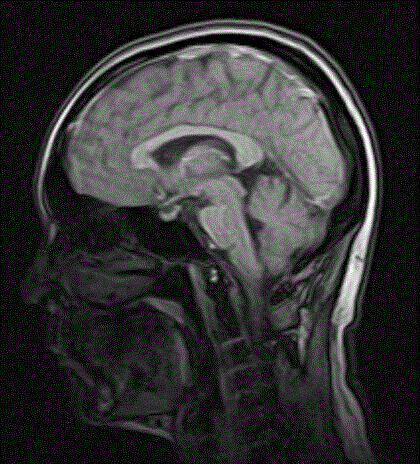
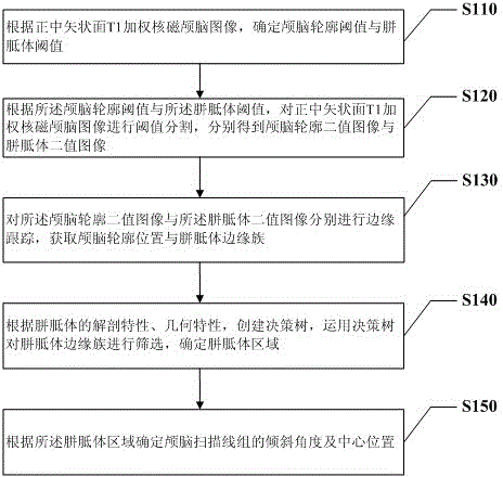
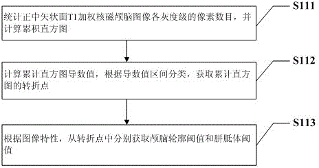
[0059] In the present invention, the T1 weighted nuclear magnetic cranial image of the median sagittal plane (such as figure 1 The corpus callosum in (shown) is automatically extracted, and the position and angle of the cranial scan line are automatically adjusted according to the extracted corpus callosum. See figure 2 , figure 2 It is a flowchart of a preferred embodiment of the method for automatically extracting the corpus callosum of a magnetic resonance image provided by the present invention. Such as figure 2 As shown, the method for automatically extracting the magnetic re...


PUM


Abstract

The invention discloses a method and system for automatically extracting the extracting magnetic resonance image corpus callosum. The method comprises the steps of according to a midsagittal plane T1 weighted nuclear magnetism brain image, determining a brain outline threshold value and a corpus callosum threshold value; performing threshold segmentation on the image according to the brain outline threshold value and the corpus callosum threshold value to respectively obtain a brain outline binary image and a corpus callosum binary image; respectively performing margin tracing on the brain outline binary image and the corpus callosum binary image to acquire a brain outline position and a corpus callosum margin cluster; establishing a decision tree, and screening marginal information to determine a corpus callosum region; determining an angle of inclination and a central position of a brain scanning line set according to the corpus callosum region. By means of the method and system, the corpus callosum in the midsagittal plane T1 weighted nuclear magnetism brain image can be extracted automatically, automatic regulation of the scanning line set during brain scanning can be completed according to information on the corpus callosum and the like, scanning efficiency is increased, no manual regulation is needed, time and labor are saved, an strong practicability is achieved.

Description

technical field [0001] The present invention relates to the technical field of magnetic resonance imaging image processing, in particular to a method and system for automatically extracting a magnetic resonance image corpus callosum. Background technique [0002] Magnetic resonance imaging (MRI for short) refers to the use of corresponding radio frequency pulses to excite the hydrogen nuclei (protons) of the subject placed in a static magnetic field, and the resulting magnetic resonance signals are collected and processed by a computer to reconstruct images. Because magnetic resonance imaging technology has the advantages of no ionizing radiation damage, high soft tissue contrast, high image resolution, flexible selection of imaging parameters and scanning orientation, and the ability to display blood vessels without contrast agents, it is widely used in clinical medical diagnosis. Its application and development The prospects are very bright. [0003] Medical image segment...

Claims


Application Information

Patent Timeline
IPC IPC(8): A61B5/055G06T7/00
Inventor 段鹏邓晓云
Owner SHENZHEN ANKE HIGH TECH CO LTD
Why Eureka
Try Eureka