
Ultrasonic diagnostic apparatus and medical image processing apparatus

A diagnostic device and ultrasonic technology, applied in the directions of acoustic wave diagnosis, infrasound wave diagnosis, image data processing, etc., can solve the problems of lack, intuition, image quality of drawn images, and realistic effects.

Active Publication Date: 2014-11-05
TOSHIBA MEDICAL SYST CORP
View PDF6 Cites 3 Cited by
  • Summary
  • Abstract
  • Description
  • Claims
  • Application Information

AI Technical Summary

Problems solved by technology

[0006] Selection of optimal rendering parameters such as light position, color map, brightness, and radiosity may have a large impact on the quality and realism of rendered images obtained from data collected by medical imaging devices
However, most operators of medical imaging devices do not have the time or expertise in rendering techniques to find and select optimal rendering parameters according to specific imaging conditions.
In addition, in the case of finding and selecting the optimal rendering parameters, the interface of some medical imaging devices is not as intuitive as the user expects

Method used


Image

  • Ultrasonic diagnostic apparatus and medical image processing apparatus
  • Ultrasonic diagnostic apparatus and medical image processing apparatus
  • Ultrasonic diagnostic apparatus and medical image processing apparatus

Examples


Embodiment Construction

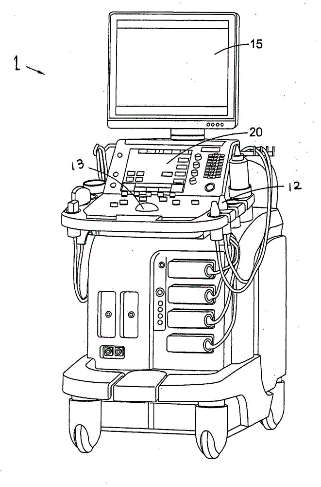
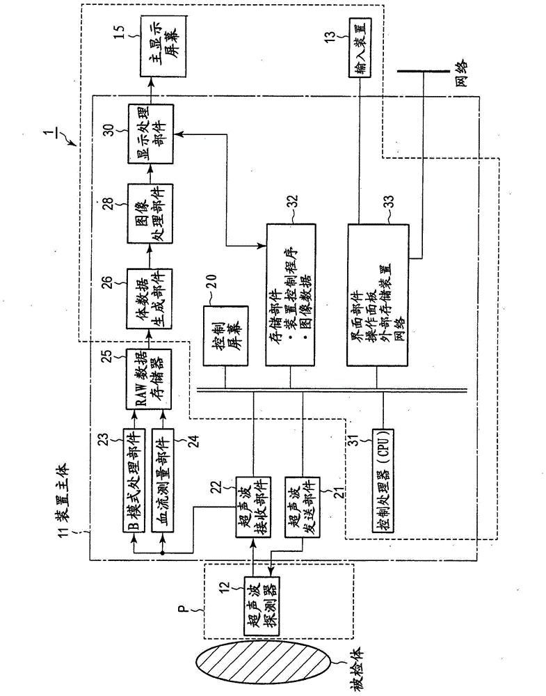
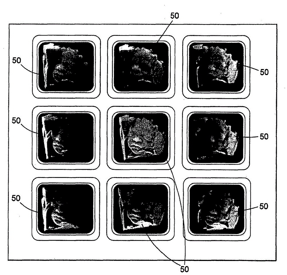
[0016] Hereinafter, an ultrasonic diagnostic apparatus according to the present embodiment will be described with reference to the drawings. In addition, in the following description, the same code|symbol is attached|subjected to the component which has substantially the same function and structure, and it repeats description only when necessary. In addition, in this embodiment, a case where a parameter adjustment function using a thumbnail image to be described later is applied to an ultrasonic image diagnosis apparatus is exemplified. However, the present invention is not limited to this example, and may be applied to other medical image diagnostic devices (for example, X-ray computed tomography devices, magnetic resonance imaging devices, X-ray diagnostic devices, etc.). In addition, imaging data acquired by various medical imaging devices may be used and applied to medical image processing devices realized by personal computers, medical workstations, and the like.

[0017...


PUM


Abstract

A medical imaging data processing apparatus comprising a data receiving unit for receiving imaging data, a rendering unit for rendering images from the imaging data according to one or more rendering parameters and a selection unit configured to output a plurality of images rendered by the rendering unit. At least one of the plurality of output images is rendered using one or more rendering parameter having a value that is different to the value used to render one or more of the other rendered images. The output images are selectable to select the value of one or more rendering parameters associated with the selected image.

Description

technical field [0001] The present embodiment relates to an ultrasonic diagnostic apparatus and a medical image processing apparatus capable of intuitively and easily changing and selecting parameters when rendering an image using data obtained by using a medical imaging apparatus typified by an ultrasonic diagnostic apparatus. Background technique [0002] As a method for improving medical visualization, generating 3D / 4D volume-rendered images from data collected by medical imaging devices has become increasingly popular. Such techniques are not limited thereto, and can be applied to various medical imaging methods such as ultrasonic imaging, MRI, CT, and PET. [0003] For example, ultrasonic imaging using transmission and reception of sound waves from a vibrator can be used to visualize internal structures. Imaging of a fetus in utero is a known example of the use of this technique. [0004] Using software, combine ultrasonic data acquired at different positions or angle...

Claims


Application Information

Patent Timeline
IPC IPC(8): A61B8/06A61B6/03A61B6/00A61B5/055G06T11/00
CPCA61B8/5207A61B8/0866A61B8/4405A61B8/465A61B8/483
Inventor M·瓦伦伯格
Owner TOSHIBA MEDICAL SYST CORP
Learn More