Eureka AIR delivers breakthrough ideas for toughest innovation challenges, trusted by R&D personnel around the world.

Mammary gland molybdenum target X-ray image block feature extraction method based on tower-shaped principal component analysis (PCA)

A feature extraction and line image technology, applied in the field of image processing, can solve the problems of incomplete use of mammary gland features, unclear definition of image block content, and no prominent importance, etc., to achieve complete density distribution features, reasonable grayscale features, and improved complete effect

Active Publication Date: 2014-12-03
XIDIAN UNIV
View PDF6 Cites 16 Cited by
  • Summary
  • Abstract
  • Description
  • Claims
  • Application Information

AI Technical Summary

Problems solved by technology

The shortcomings of this method are: the feature dimension is too high, there is information redundancy, the importance of the overall image block with high density in the middle and low density at the edge is not highlighted, and the classification accuracy is not high.
The disadvantage of this method is that the division method of the image block does not clearly define the content contained in the image block, and cannot be fully used in breast features.

Method used


Image

  • Mammary gland molybdenum target X-ray image block feature extraction method based on tower-shaped principal component analysis (PCA)
  • Mammary gland molybdenum target X-ray image block feature extraction method based on tower-shaped principal component analysis (PCA)
  • Mammary gland molybdenum target X-ray image block feature extraction method based on tower-shaped principal component analysis (PCA)

Examples


Embodiment Construction

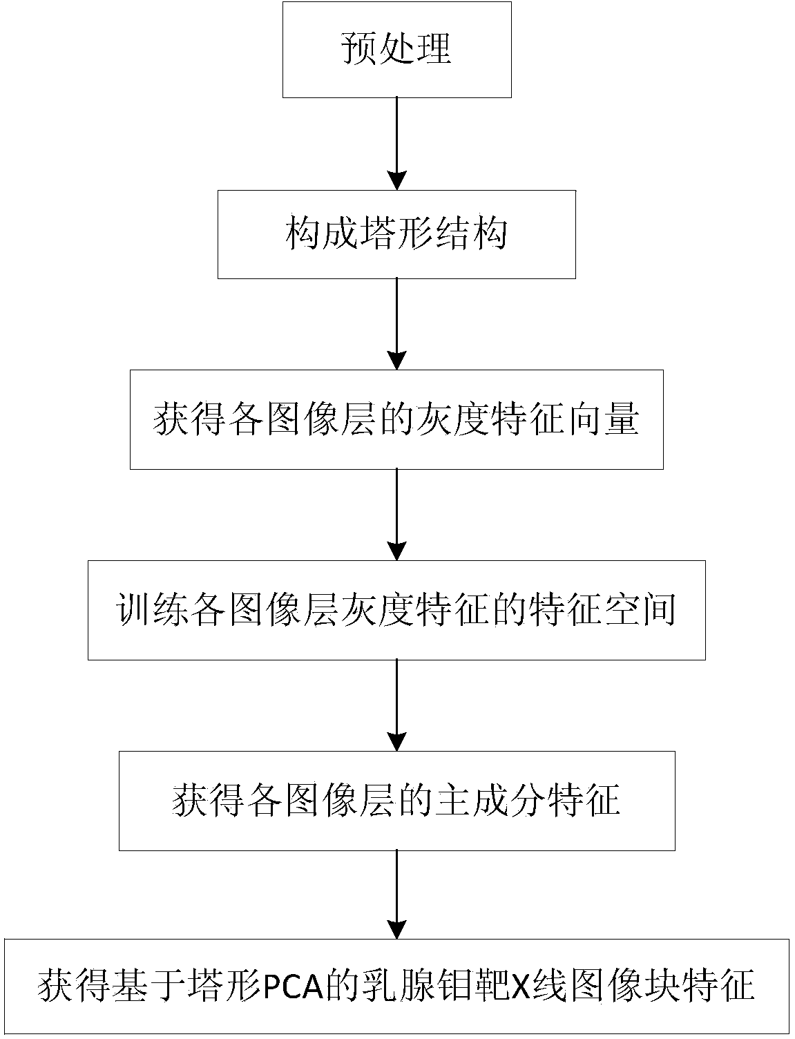
[0043] Attached below figure 1 , further describe in detail the steps realized by the present invention.

[0044] Step 1, preprocessing.

[0045] The mammography image is preprocessed, and the mammography image block is obtained through the sliding window. The width of the mammography image block is 100 pixels, and the height of the mammography image block is 100 pixels.

[0046] The method for preprocessing the mammography image is carried out as follows:

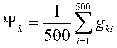
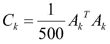
[0047] The first step is to use the median filter method to denoise the mammography images: set the sliding window of the median filter to a square window of 3×3 pixels, and use the square window along the mammogram The direction of the X-ray photographic image line slides pixel by pixel. During each sliding period, the gray values ​​of all pixels in the square window are sorted in ascending order, and the middle value of the sorting result is selected to replace the square window. The gray value of the pixel at the ce...


PUM


Abstract

The invention discloses a mammary gland molybdenum target X-ray image block feature extraction method based on tower-shaped principal component analysis (PCA). The mammary gland molybdenum target X-ray image block feature extraction method based on tower-shaped PCA mainly overcomes the defect that features extracted in the prior art do not contain the feature that the density of the middle of a lump is large while the density of the edge of the lump is small. The method comprises the following steps of (1) carrying out pretreatment, (2) constituting a tower-shaped structure, (3) obtaining a gray feature vector of each image layer, (4) training a feature space of the gray feature of each image layer, (5) obtaining principal component features of each image layer, and (6) obtaining mammary gland molybdenum target X-ray image block features based on tower-shaped PCA. According to the method, the mammary gland molybdenum target X-ray image block features can be represented more robustly, image features can be represented more effectively, the accurate rate of detection of a lump region in a mammary gland molybdenum target X-ray photography image is increased, and therefore radiologists are assisted to carry out clinical diagnosis.

Description

technical field [0001] The invention belongs to the technical field of image processing. It further relates to a mammography X-ray image block grayscale feature extraction method based on tower-shaped principal component analysis (PCA) in the field of medical image processing technology. According to the distribution of mammary gland mass images with high density in the middle and low density at the edges, the present invention extracts layered features of breast image blocks, and at the same time performs principal component analysis on the gray features extracted by layers, thereby improving mammography images. Accuracy of mass region detection. The invention can be used for detection of lesion areas in clinical medicine, improves the detection rate, reduces the detection false positive rate, and assists radiologists in clinical diagnosis. Background technique [0002] At present, the image features used in clinical medical diagnosis are gray-scale intuitive features, gr...

Claims


Application Information

Patent Timeline
Patent Type & Authority Applications(China)
IPC IPC(8): G06K9/46G06T7/00A61B6/03
Inventor 李洁王颖刘璐高锐逄敏焦志成王斌路文李圣喜张琪
Owner XIDIAN UNIV
Why Eureka
Eureka Blog
Learn More