
A kind of ct scan image reconstruction method and ct scanner

A technology of image reconstruction and CT scanning, applied in the field of medical devices, can solve the problems of abnormal CT value, truncation of projection data, inability to obtain projection data, etc., to overcome technical shortcomings, improve accuracy, and eliminate the possibility of truncation artifacts. Effect

Active Publication Date: 2017-08-25
NEUSOFT MEDICAL SYST CO LTD
View PDF7 Cites 0 Cited by
  • Summary
  • Abstract
  • Description
  • Claims
  • Application Information

AI Technical Summary

Problems solved by technology

This method reduces the X-rays irradiated to the human body and saves the X-ray dose, but at the same time it causes a problem: figure 1 The projection data of the area outside the circle of radius R' in the right figure cannot be obtained, that is, the projection data is truncated
The appearance of truncation artifacts will lead to image distortion and abnormal CT values, which may mislead doctors in the diagnosis of diseases and affect the recovery of patients.

Method used


Image

  • A kind of ct scan image reconstruction method and ct scanner
  • A kind of ct scan image reconstruction method and ct scanner
  • A kind of ct scan image reconstruction method and ct scanner

Examples


Embodiment 1

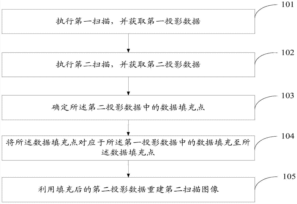
[0069] see figure 2 , which is a flow chart of Embodiment 1 of a CT scan image reconstruction method provided by the present invention.

[0070] The CT scanning image reconstruction method provided by the embodiment of the present invention comprises the following steps:

[0071] Step 101: Execute a first scan, the first scan is a scan of a complete human body in a tomographic plane, and acquire first projection data.

[0072] After the doctor confirms the patient's information, scheduled scanning location, and patient's position on the scanning bed, he usually performs some scans of the entire human body in the tomographic plane, such as human body positioning scans, contrast agent positioning scans, etc. At this time, the The slit width of the collimator reaches the width that X-rays can cover the complete human body in the tomographic plane, for example figure 1 left image in . Among them, the human body positioning scan is used to locate the position of the patient on ...

Embodiment 2

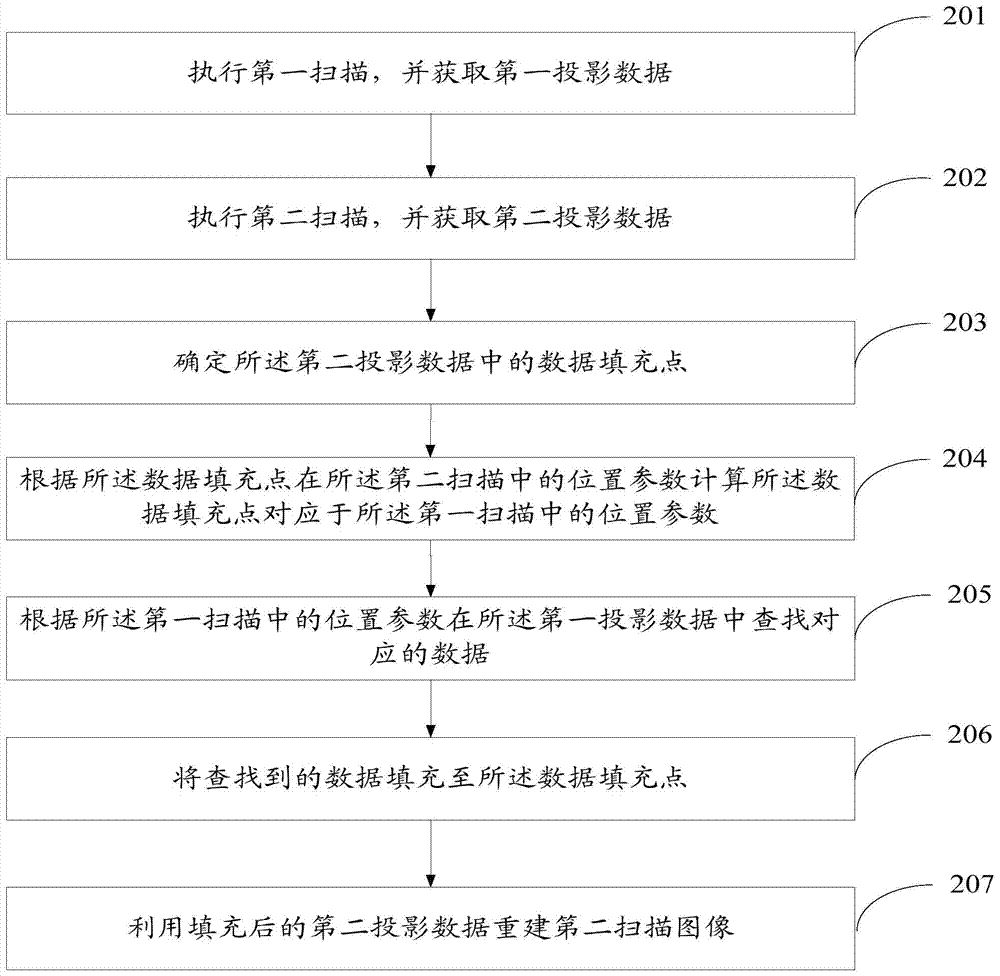
[0088] see image 3 , which is a flow chart of Embodiment 2 of a CT scan image reconstruction method provided by the present invention.

[0089] The CT scanning image reconstruction method provided by the embodiment of the present invention comprises the following steps:

[0090] Step 201: Execute a first scan, which is a scan of a complete human body in a tomographic plane, and acquire first projection data.

[0091] Step 202: Execute a second scan, the second scan is a scan of a part of the human body within a tomographic plane, and acquire second projection data.

[0092] Step 203: Determine a data filling point in the second projection data, wherein the data filling point is located in a truncated area.

[0093] Step 204: Calculate the position parameter corresponding to the data filling point in the first scan according to the position parameter of the data filling point in the second scan.


PUM


Abstract

The embodiment of the present invention discloses a CT scan image reconstruction method, the method comprising: performing a first scan and obtaining first projection data; performing a second scan and obtaining second projection data; determining the second projection data fill the data in the data filling point; fill the data filling point corresponding to the data in the first projection data to the data filling point; use the filled second projection data to reconstruct the second scanning image. The embodiment also discloses a CT scanner. The invention eliminates the truncation artifact of the reproduced image, improves the quality of the reconstructed image, and increases the accuracy of the doctor's disease diagnosis.

Description

technical field [0001] The invention relates to the field of medical equipment, in particular to a CT scan image reconstruction method and a CT scanner. Background technique [0002] CT (Computed Tomography) is electronic computerized tomography. During the clinical CT scanning process, it often includes multiple sets of scanning sequences. sequence etc. In the traditional technology, in order to obtain complete projection data without truncation, the scanned X-ray needs to cover the whole body of the patient, see figure 1 , figure 1 The left and right diagrams are cross-sectional views perpendicular to the line connecting the head and feet of the human body, that is, cross-sectional views perpendicular to the direction of entering the bed. The X-ray source 102 irradiates the scanning bed 103 through the gap of the collimator 101 . The X-ray source refers to a device that generates X-rays, the collimator refers to a component that generates a radiation field of a certain ...

Claims


Application Information

Patent Timeline
Patent Type & Authority Patents(China)
IPC IPC(8): A61B6/03G06T11/00G16H30/20G16H30/40
CPCA61B6/488A61B6/032G16H30/40G16H30/20A61B6/52G16H20/40
Inventor 楼珊珊郑晗
Owner NEUSOFT MEDICAL SYST CO LTD
Learn More