
High-flux shooting method used for flow cytometry imaging
A technology of flow cytometer and shooting method, which is applied in the direction of image communication, color TV parts, TV system parts, etc., can solve the problems of low shooting efficiency, low luminous flux, missing shots, etc., to improve shooting efficiency, The effect of increasing luminous flux
- Summary
- Abstract
- Description
- Claims
- Application Information
AI Technical Summary
Problems solved by technology
Method used
Image

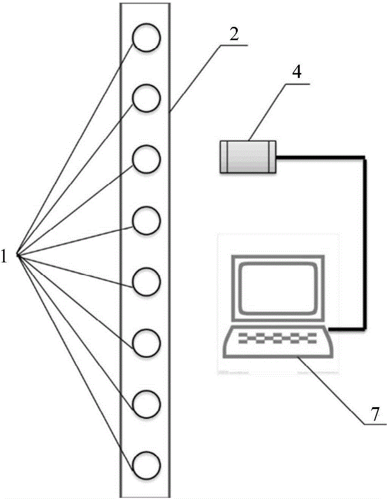
Examples
Embodiment 1
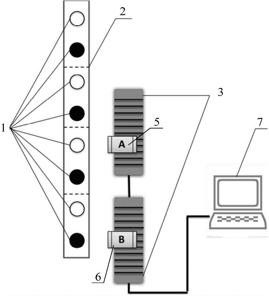
[0031] See attached figure 2 , the device used to realize the high-throughput shooting method for imaging flow cytometer of the present invention includes micropipe 2, transmission device 3, camera A5, camera B6 and computer 7; said camera A5 and camera B6 respectively The tracking and shooting of the cells in the micro-channel 2 is realized by driving the transmission device 3 , and the movement of the transmission device 3 is controlled by a computer 7 .
[0032] A high-throughput shooting method for imaging flow cytometer of the present invention comprises the following steps:
[0033] Step 1: Cell 1 moves from top to bottom from microchannel 2, the flow rate of cell 1 is 2V, and the cells in microchannel 2 are sequentially numbered as n, n+1, n+2...n+m from bottom to top;
[0034] Step 2: When the n+1th cell enters the field of view of the camera A5, the camera A5 starts to work and takes pictures of the n+1th cell, and the camera A5 drives the n+1th cell by △V through t...
Embodiment 2
[0040] Cell 1 flows from top to bottom along the microchannel 2, we divide two adjacent cells 1 into a group, see attached figure 2, draw four groups of cells respectively, numbered as group 1 to group 4 from bottom to top according to the flow order of cell 1. And cell 1 is numbered from bottom to top, respectively: 1 group n, n+1, 2 groups n+2, n+3, 3 groups n+4, n+5 and 4 groups n+6, n+7 The cells in group 1 flow through camera A5 first, and then through camera B6. When cell n+1 in group 1 enters the field of view of camera A5, camera A5 starts to work, and camera A5 follows the cells under the action of transmission device 3. The flow of n+1 moves in the same direction to complete the exposure imaging of cell n+1 in the field of view of camera A for △t time, and then camera A5 quickly resets, waiting for the next group of cell 1 to be photographed. Then cell n of group 1 enters the field of view of camera B6, and cell n+3 of group 2 enters the field of view of camera A5....
PUM
 Abstract
Description
Claims
Application Information