
Devices and Methods for Perfusing an Organ

a technology for supporting devices and organs, applied in the field of supporting devices, can solve the problems of reduced pressure in the vessel, deformation of the vessel wall, and complete obstruction of the vessel, and achieve the effects of preventing collision of collection catheters, reducing flow rates, and maintaining patency of the vessel

Inactive Publication Date: 2009-01-15
OSPREY MEDICAL
View PDF28 Cites 121 Cited by
  • Summary
  • Abstract
  • Description
  • Claims
  • Application Information

AI Technical Summary

Benefits of technology

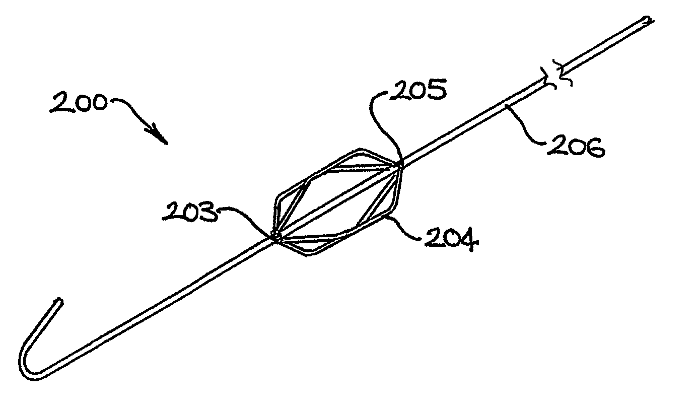
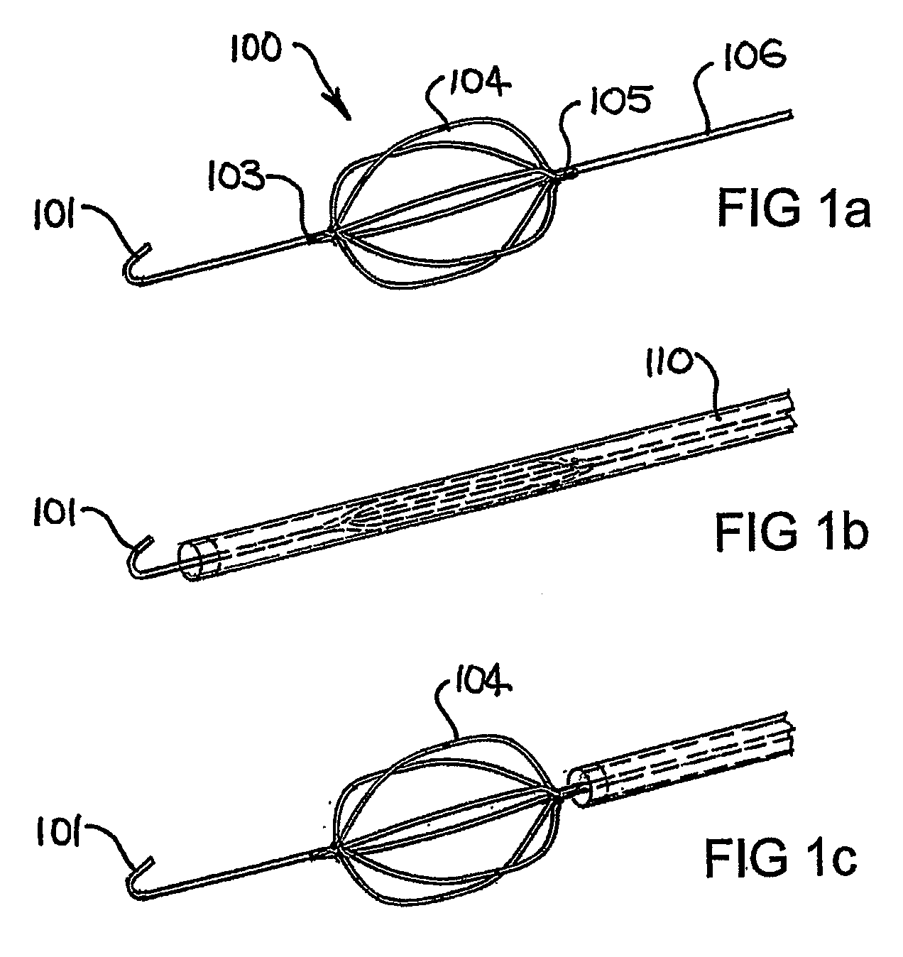
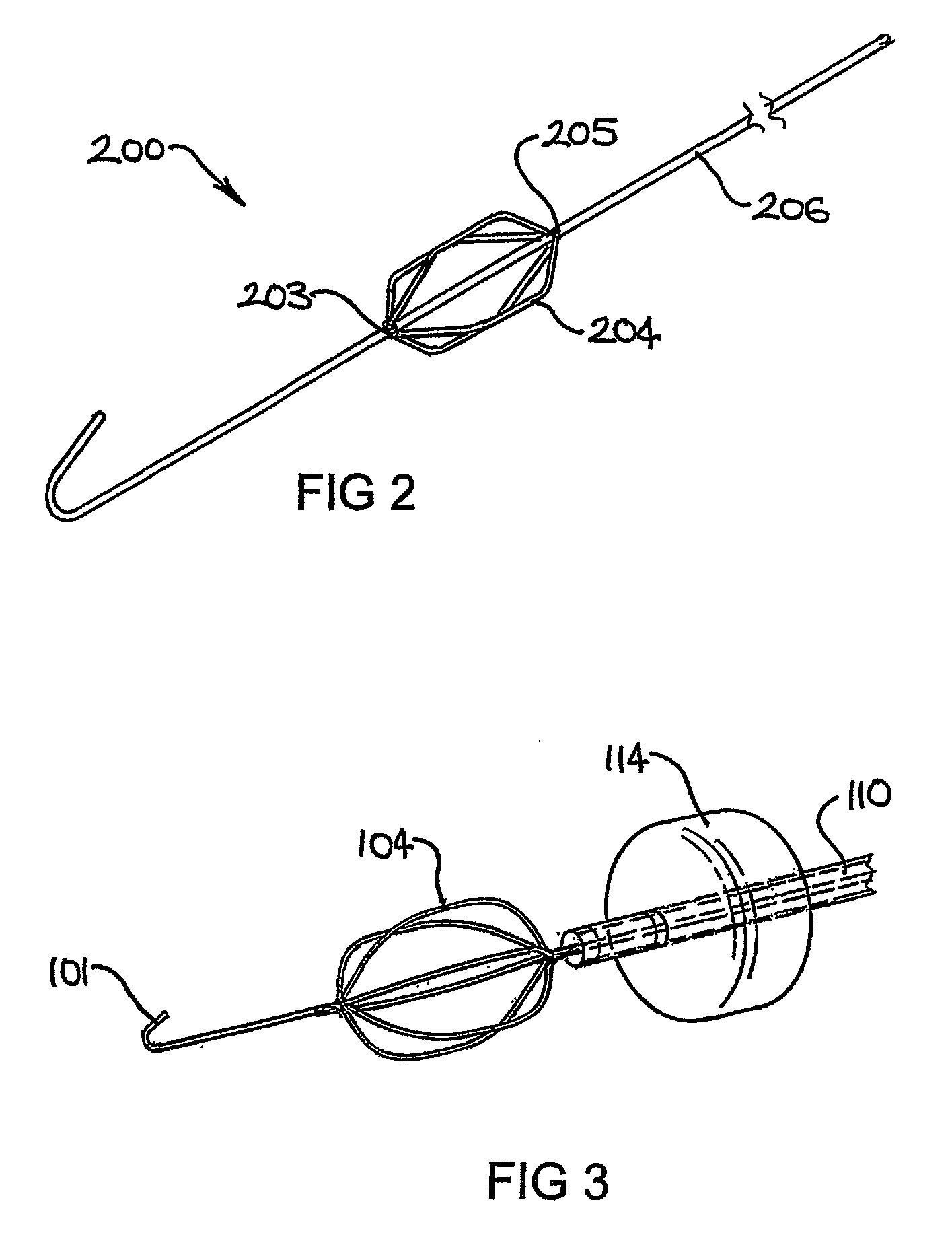
[0010]In one aspect the present invention provides a percutaneously deliverable device for supporting a vessel in a human or animal subject including means for supporting the vessel during delivery of a fluid thereto or collection of fluid therefrom. Without wishing to be limited by theory, Applicant proposes that collapse of the vessel wall is at least partly responsible for the low flow rates that are problematic in the percutaneous collection of fluids from an organ or tissue of the body. The support devices described herein have the effect of maintaining patency of the vessel via which fluid is being collected, while also preventing collision of the collection catheter with the vessel wall. Maintenance of patency of the collection vessel allows for useful flow rates to be established, thereby reducing the risk of venous pooling. Where the invention further includes delivery of a fluid to a tissue or organ, flow rates of the fluid into the tissue or organ are also improved.

Problems solved by technology

Movement of fluids into and out of a vessel can cause deformations in the wall of the vessel leading to collapse.
The collapse of the vessel wall can lead to complete obstruction of the vessel thereby preventing any further flow of fluid.
In particular drawing fluid out of a vessel during collection can lead to a decrease in pressure in the vessel which in turn leads to vessel collapse.
Under typical perfusion conditions, the rate at which fluid is drawn out of the vessel may need to be sufficiently high such that the vessel walls cannot maintain patency and subsequently collapse.
The cause of vessel collapse may be due to a combination of high flow rates and low pressure on the collection side of the organ.
A particularly problematic case is where the coronary sinus is used to collect fluid perfusing through the coronary circulation of the heart.
The coronary sinus can be difficult to deal with when collecting fluid using a collection lumen located within the sinus.
This is in part due to the fact that the coronary sinus wall is flaccid and prone to collapse when contacted by a collection catheter tip.
This problem is intensified as a result of the low pressures which are generated at the catheter tip as fluid is drawn out of the sinus and these low pressures, in combination with inadequate flow into the sinus, can cause the vessel wall to collapse.

Method used


Image

  • Devices and Methods for Perfusing an Organ
  • Devices and Methods for Perfusing an Organ
  • Devices and Methods for Perfusing an Organ

Examples


example 1

Effect of Support Device on Flow Rates and Pressures Achievable During Recirculation in Sheep Right Hepatic Vein, Cephalic Vein, Coronary Sinus and Renal Vein During Recirculation

[0105]A 0.014″ diameter superelastic nitinol wire stem of 1.35 m length was used, coupled to an expandable member having 6 pre-shaped elliptical loop portions welded to the stem. A 0.024″ OD atraumatic tip of 2 cm length attached to the distal end of the expandable member was used to position the device in the blood vessel. A balloon occlusion catheter was positioned in the vessel and the expandable member deployed at the tip of the catheter. The balloon was inflated to isolate and capture flows in the vessel and the catheter was connected to a standard extracorporeal circuit for blood circulation.

TABLE 1Perfusion of the right hepatic and cephalic veins.pressure (mmHg)Flowright hepatic veincephalic vein(mL / min)deviceno devicedeviceno device20−20040−10−13−11↓60−20−19−18no flow80−30−23achievable100−30−47−3212...


PUM


Abstract

The present invention provides devices and methods for use in the perfusion of organs and anatomical regions. In one aspect the present method provides a percutaneously deliverable device for supporting a vessel in a human or animal subject including means for supporting the vessel during delivery of a fluid thereto or collection of a fluid therefrom. In another aspect the invention provides a method for delivery or collection of a fluid to or from an organ or anatomical region in a human or animal subject, the method including the step of supporting a vessel associated with the organ or anatomical region. The devices and methods may be used to deliver, remove or recirculate a therapeutic agent to an organ or anatomical region.

Description

FIELD OF THE INVENTION[0001]The present invention relates to a support device for supporting a vessel of a human or animal subject. The support device can be used, for example, during procedures in which a fluid is collected from an organ or anatomical region via an associated blood vessel.BACKGROUND TO THE INVENTION[0002]Many therapeutic procedures can be performed percutaneously. Such procedures include investigative and diagnostic methods as well as medical and surgical treatments. Percutaneous techniques are advantageous because they are minimally invasive, and therefore present a reduced risk to the patient when compared with invasive surgeries requiring open access to tissues within the patient's body.[0003]Catheters and sheaths are typically used to access internal sites by entering the patient's body through an incision or other entry point to the peripheral vasculature and navigating through the torturous paths of the vascular system to the target site. Tools, cameras illum...

Claims


Application Information

Patent Timeline
Patent Type & Authority Applications(United States)
IPC IPC(8): A61M25/01A61F2/84
CPCA61M25/09A61M29/02A61F2/82A61M2025/1052A61M2025/09183A61M25/01A61M25/10
Inventor POWER, JOHN MELMOUTHMATHIS, MARK L.KAYE, DAVID MARTINBILNEY, ADAM LUCAS
Owner OSPREY MEDICAL
Learn More