Devices for maintaining surgically created openings

a technology of surgical creation and openings, applied in the direction of surgical staples, prostheses, blood vessels, etc., can solve the problems of copd-afflicted individuals, loss of muscle strength, and inability to perform common daily activities, so as to prevent additional wound healing and reduce tissue growth

Inactive Publication Date: 2005-06-23
BIGGS MICHAEL +12
View PDF99 Cites 104 Cited by
  • Summary
  • Abstract
  • Description
  • Claims
  • Application Information

AI Technical Summary

Benefits of technology

[0025] The devices and methods described herein also serve to maintain the patency of a channel surgically created in an airway wall. In particular, the methods and devices prevent closure of the channel such that air may flow through the channel and into the airway. The step of preventing closure of the airway may be performed a number of ways including (1.) impeding the wound healing process of the lung tissue such that the lung tissue cannot heal and the channel remains patent; or (2.) accelerating the wound healing process such that the channel remains patent. Accelerating the wound healing process may be carried out, for example, by increasing the growth of epithelial cells.
[0029] Substances which are known to prevent infection may also be used in the present invention. Antibiotics, for example, and other infection-fighting substances can serve to prevent additional wound healing processes which normally commence when an infection or bacteria is present at a wound or injury site.
[0030] Conduits for maintaining the patency of a channel created in tissue may comprise a radially expandable center section having a first end and a second end and a passageway extending between the ends. The conduit may further include at least one center-control segment configured to restrict radial expansion of the passageway to a maximum profile. At least one extension member may extend from each of the first and second ends of the center section and each of the extension members may have a fixed end connected to one of the ends of the center section and a movable end such that each of the extension members is capable of being deflected about the fixed end. The conduit further includes a bioactive substance disposed on at least a portion of a surface of the conduit. The bioactive substance may serve to reduce tissue growth such that the conduit remains in the channel and the passageway remains at least partially open. The bioactive substance may be disposed on regions of the surface corresponding to the center section, the extension members, both the center section and extension members, or portions of these features.
[0034] In another variation of the present invention, the conduit includes at least one hold-down member extending from the tips (or another location) of the deflecting members. The hold-down members serve to prevent the conduit from being ejected. The hold-down members desirably include one or more regions which are susceptible to tissue ingrowth or overgrowth. In some embodiments of the present invention, the hold-down members include spaces for tissue to grow into such that it may reconnect with itself, encapsulating the hold-down member and thus preventing ejection of the conduit.
[0036] The conduit may comprise at least one visualization feature disposed on a portion of the tissue barrier. The visualization feature may be a stripe circumferentially disposed about at least a portion of the center section or it may be disposed on the extension members or the hold-down members. The visualization feature serves to aid in placement or deployment of the conduit in a target site.

Problems solved by technology

Those inflicted with COPD face disabilities due to the limited pulmonary functions.
Usually, individuals afflicted by COPD also face loss in muscle strength and an inability to perform common daily activities.
Since the damage to the lungs is irreversible, there is little hope of recovery.
Most times, the physician cannot reverse the effects of the disease but can only offer treatment and advice to halt the progression of the disease.
However, these conducting airways do not take part in gas exchange because they do not contain alveoli.
If the lungs' ability to recoil is damaged, the lungs cannot contract and reduce in size from their inflated state.
As a result, the lungs cannot evacuate all of the inspired air.
The destruction of the alveolar walls results in a dual problem of reduction of elastic recoil and the loss of tethering of the airways.
Unfortunately for the individual suffering from emphysema, these two problems combine to result in extreme hyperinflation (air trapping) of the lung and an inability of the person to exhale.
In this situation, the individual will be debilitated since the lungs are unable to perform gas exchange at a satisfactory rate.
While alveolar wall destruction decreases resistance to collateral ventilation, the resulting increased collateral ventilation does not benefit the individual since air is still unable to flow into and out of the lungs.
Yet, bronchodilator drugs are only effective for a short period of time and require repeated application.
Moreover, the bronchodilator drugs are only effective in a certain percentage of the population of those diagnosed with COPD.
Unfortunately, aside from the impracticalities of needing to maintain and transport a source of oxygen for everyday activities, the oxygen is only partially functional and does not eliminate the effects of the COPD.
Moreover, patients requiring a supplemental source of oxygen are usually never able to return to functioning without the oxygen.
However, lung reduction surgery is an extremely traumatic procedure which involves opening the chest and thoracic cavity to remove a portion of the lung.
If the entire lung is emphysematous, however, removal of a portion of the lung removes gas exchanging alveolar surfaces, reducing the overall efficiency of the lung.
Lung volume reduction surgery is thus not a practical solution for treatment of emphysema where the entire lung is diseased.
Both bronchodilator drugs and lung reduction surgery fail to capitalize on the increased collateral ventilation taking place in the diseased lung.
Events that may arise when a device is implanted in a surgically-created channel in a lung is that the device can be ejected, filled in with tissue, or otherwise rendered ineffective as the wound heals.

Method used


Image

  • Devices for maintaining surgically created openings
  • Devices for maintaining surgically created openings
  • Devices for maintaining surgically created openings

Examples


Embodiment Construction

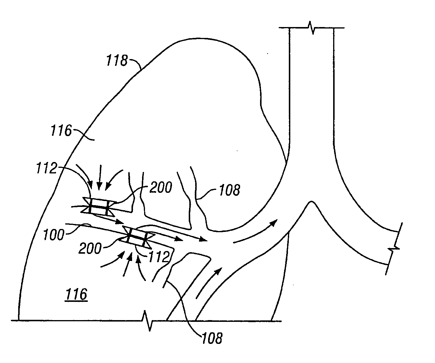
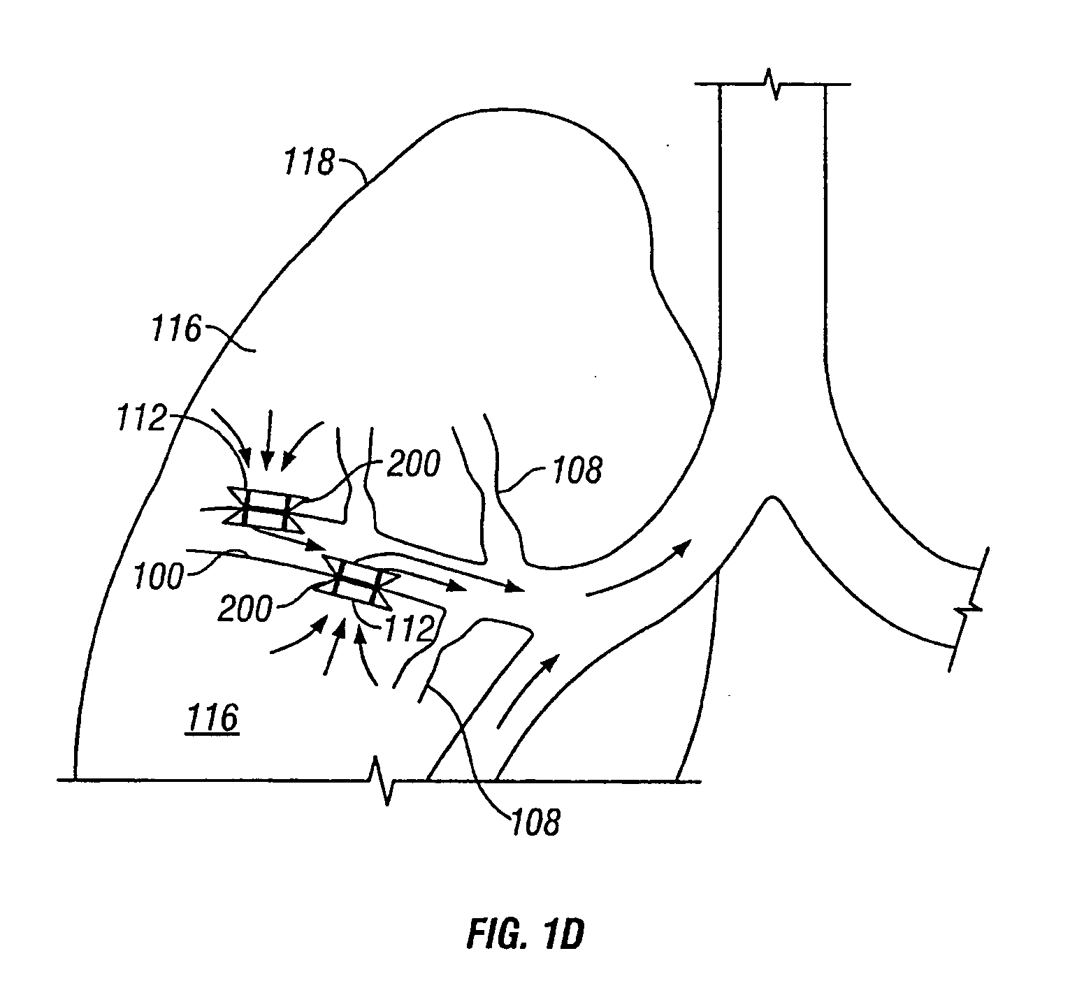
[0062] Described herein are devices and methods for improving the gaseous exchange in the lung. In particular, a conduit is described that serves to maintain collateral openings or channels surgically created through an airway wall so that air is able to pass directly out of the lung tissue and into the airways. This facilitates exchange of oxygen into the blood and decompresses hyper inflated lungs.

[0063] By “channel” it is meant to include, but not be limited to, any opening, hole, slit, channel or passage created in the airway wall. The channel may be created in tissue having a discrete wall thickness and the channel may extend all the way through the wall. Also, a channel may extend through lung tissue which does not have well defined boundaries such as, for example, parenchymal tissue.

[0064] As stated above, the conduits described herein may improve airflow through an airway in the lung. Simplified illustrations of various states of a natural airway and a blood gas interface ...


PUM


Abstract

Devices and methods are directed to improving the gaseous exchange in a lung of an individual having, for instance, chronic obstructive pulmonary disease. More particularly, conduits may be deployed in the lung to maintain collateral openings (or channels) surgically created through airway walls. This tends to facilitate both the exchange of oxygen ultimately into the blood and decompress hyper-inflated lungs.

Description

CROSS REFERENCE TO RELATED APPLICATIONS [0001] This is a continuation of U.S. patent application Ser. No. 10 / 951,962 filed on Sep. 28, 2004 which is a continuation of international application No. PCT / US03 / 12323 filed on Apr. 21, 2003, which is a non-provisional of U.S. provisional patent application No. 60 / 374,022 filed on Apr. 19, 2002, and a non-provisional of U.S. provisional patent application No. 60 / 387,163 filed on Jun. 7, 2002, and a non-provisional of U.S. provisional patent application No. 60 / 393,629 filed on Jul. 3, 2002. The entirety of all of the above listed applications are hereby incorporated by reference.FIELD OF THE INVENTION [0002] The invention relates to devices and methods for improving the gaseous exchange in a lung of an individual having, for instance, chronic obstructive pulmonary disease. More particularly, the invention relates to conduits that are deployed in the lung to maintain collateral openings (or channels) surgically created through an airway wall...

Claims


Application Information

Patent Timeline
Patent Type & Authority Applications(United States)
IPC IPC(8): A61F2/02A61B17/00A61B17/02A61B17/064A61B17/068A61B17/12A61F2/04A61F2/86
CPCA61B17/0218A61F2230/0078A61B17/068A61B17/12022A61B17/12104A61B17/12172A61B2017/00252A61B2017/0046A61B2017/00477A61F2/07A61F2/2412A61F2/86A61F2/91A61F2002/043A61F2230/0054A61F2220/0008A61F2220/0016A61F2230/005A61B17/064
Inventor BIGGS, MICHAELKEAST, THOMASLOOMAS, BRYANTANAKA, DONTHOMPSON, DAVIDKAPLAN, GARYSHRINER, KELLYKARABEY, HALILREDMOND, RUSSVIDAL, CLAUDECOLLINSON, MIKECOLE, CARYWILLINK, MICHAEL
Owner BIGGS MICHAEL
Why Eureka
Try Eureka