Arteriovenous retinal vessel optic disk positioning method of eye fundus image

A technology of retinal blood vessels and fundus images, which is applied in the fields of eye testing equipment, medical science, diagnosis, etc., can solve the problems of low degree of automation, achieve robustness and reduce the effect of rejection

Active Publication Date: 2015-04-29
海宁求是创新健康科技有限公司
View PDF4 Cites 9 Cited by
  • Summary
  • Abstract
  • Description
  • Claims
  • Application Information

AI Technical Summary

Problems solved by technology

[0007] Blood vessel segmentation, optic disc positioning and vessel classification (arteriovenous split) on fundus images are the basis of retinal v...

Method used


Image

  • Arteriovenous retinal vessel optic disk positioning method of eye fundus image
  • Arteriovenous retinal vessel optic disk positioning method of eye fundus image
  • Arteriovenous retinal vessel optic disk positioning method of eye fundus image

Examples


Embodiment Construction

[0032] The present invention will be described in detail below with reference to the drawings and specific embodiments.

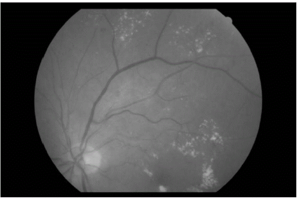
[0033] This embodiment takes figure 1 The fundus image shown is taken as an example to illustrate the method for locating the arteriovenous, retinal, and optic discs of the fundus image, and the size of the fundus image is 3000×3000. There are bright rings in the fundus image due to the ring reflection caused by photography, the non-vascular step edge around the optic disc, patchy lesions, and hemorrhagic lesions.

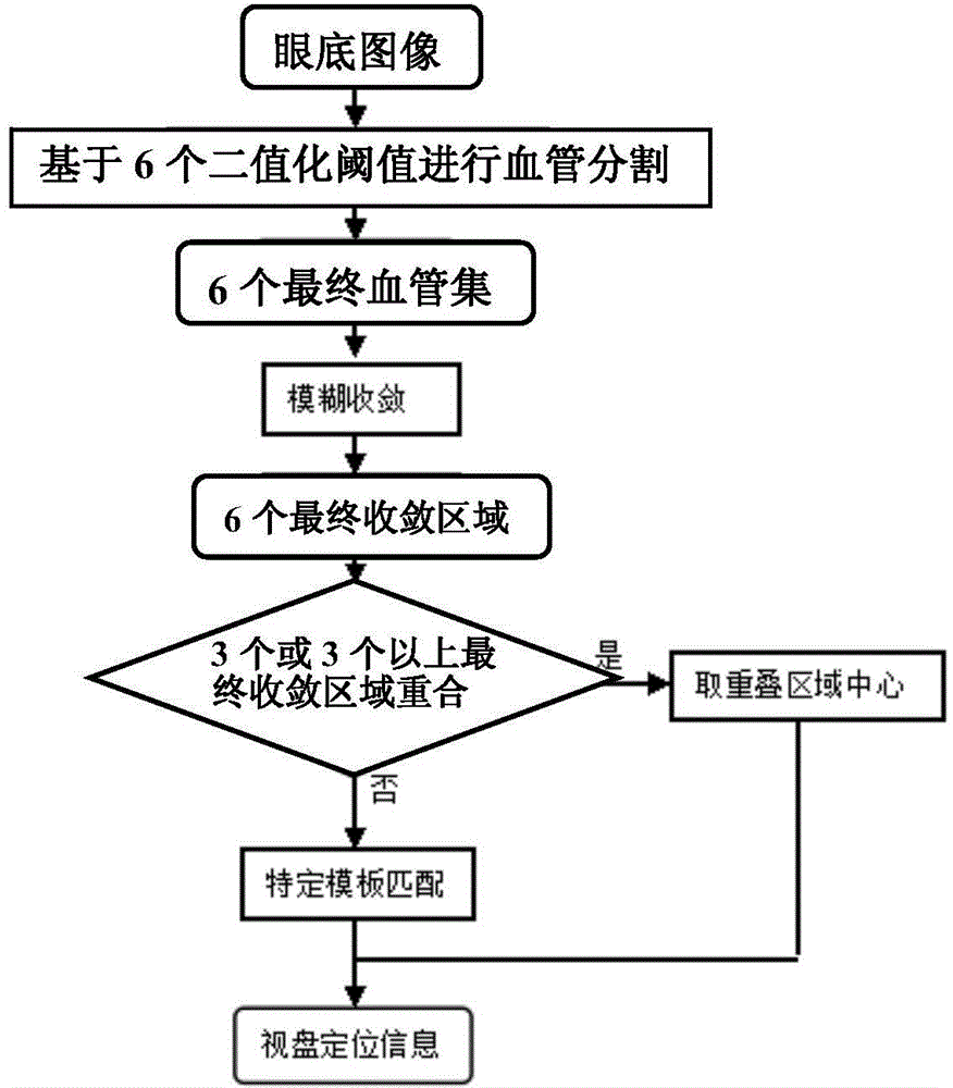
[0034] Based on the fundus image, the fundus image based on the breadth search algorithm is used to locate the optic disc of the arteriovenous retina. The process of optic disc positioning is as follows: figure 2 shown, including the following steps:

[0035] (1) Obtain the global blood vessel set (i.e. the final blood vessel set) of the fundus image, and the global blood vessel set is the collection of all blood vessels in the fundus image; ...


PUM


Abstract

The invention discloses an arteriovenous retinal vessel optic disk positioning method of an eye fundus image. The method comprises the following steps: acquiring a global vessel set of the eye fundus image, wherein the global vessel set is a set of all vessels in the eye fundus image; acquiring a convergence region of each vessel by using a fuzzy convergence algorithm aiming at each vessel in the global vessel set; counting the number of the convergence regions to which each pixel point of the eye fundus image belongs to serve as voting value of the pixel point; selecting the previous n pixel points with large voting values, applying an eight connection-based region communication algorithm to the selected n pixel points to obtain a plurality of communication regions, and taking a central coordinate of the communication region with the maximum area as the optic disk positioning information. According to the optic disk positioning method, a multiple positioning voting mode is adopted, the method has robustness and behaves well under the complex condition of more diseases in the eye fundus image, finally a template is added for matching to serve as positioning and non-convergent correction, and the rejection is less compared with the method that the condition is not treated.

Description

technical field [0001] The invention relates to the technical field of computer-aided diagnosis, in particular to a method for locating arteriovenous, retinal, and optic discs of fundus images. Background technique [0002] With the rapid development of the field of artificial intelligence in computer technology, computer-aided diagnosis technology is also gradually developed. Computer-aided diagnosis technology refers to the use of imaging, medical image processing technology and other possible physiological and biochemical means, combined with computer analysis and calculation, to assist radiologists in finding lesions and improving the accuracy of diagnosis. [0003] Usually, computer-aided diagnosis in medical imaging is divided into three steps, as follows: the first step is to extract the lesion from the normal structure; the second step is to quantify the image features; the third step is to process the data and draw conclusions . [0004] Because the computer can f...

Claims


Application Information

Patent Timeline
IPC IPC(8): A61B3/15
Inventor 吴健黎罗河邓水光李莹尹建伟吴朝晖
Owner 海宁求是创新健康科技有限公司
Why Eureka
Try Eureka