A multi-mode hysteroscopy system and its realization method

An implementation method and hysteroscopy technology, applied in the field of endoscopy, can solve the problems of limited tissue penetration depth, insufficient diagnostic accuracy, and inability to examine deep-level tissues, and achieve the effect of simplifying the scanning probe and facilitating production.

Active Publication Date: 2016-04-20
GUANGDONG OPTO MEDIC TECH CO LTD
View PDF5 Cites 0 Cited by
  • Summary
  • Abstract
  • Description
  • Claims
  • Application Information

AI Technical Summary

Problems solved by technology

[0004] However, because early uterine cancer often occurs below the mucosal layer, although the basic shape of the uterine mucosal epithelium can be obtained by cervical scope, due to the use of white light illumination, the penetration depth of white light to the tissue is limited, and the deep tissue cannot be inspected. lack of accuracy
At present, there is no instrument for deep imaging diagnosis of the fine structure of uterine mucosa

Method used


Image

  • A multi-mode hysteroscopy system and its realization method
  • A multi-mode hysteroscopy system and its realization method
  • A multi-mode hysteroscopy system and its realization method

Examples


Embodiment Construction

[0033] The present invention provides a multi-mode hysteroscopy system and its implementation method. In order to make the purpose, technical solution and effect of the present invention clearer and clearer, the present invention will be further described in detail below. It should be understood that the specific embodiments described here are only used to explain the present invention, not to limit the present invention.

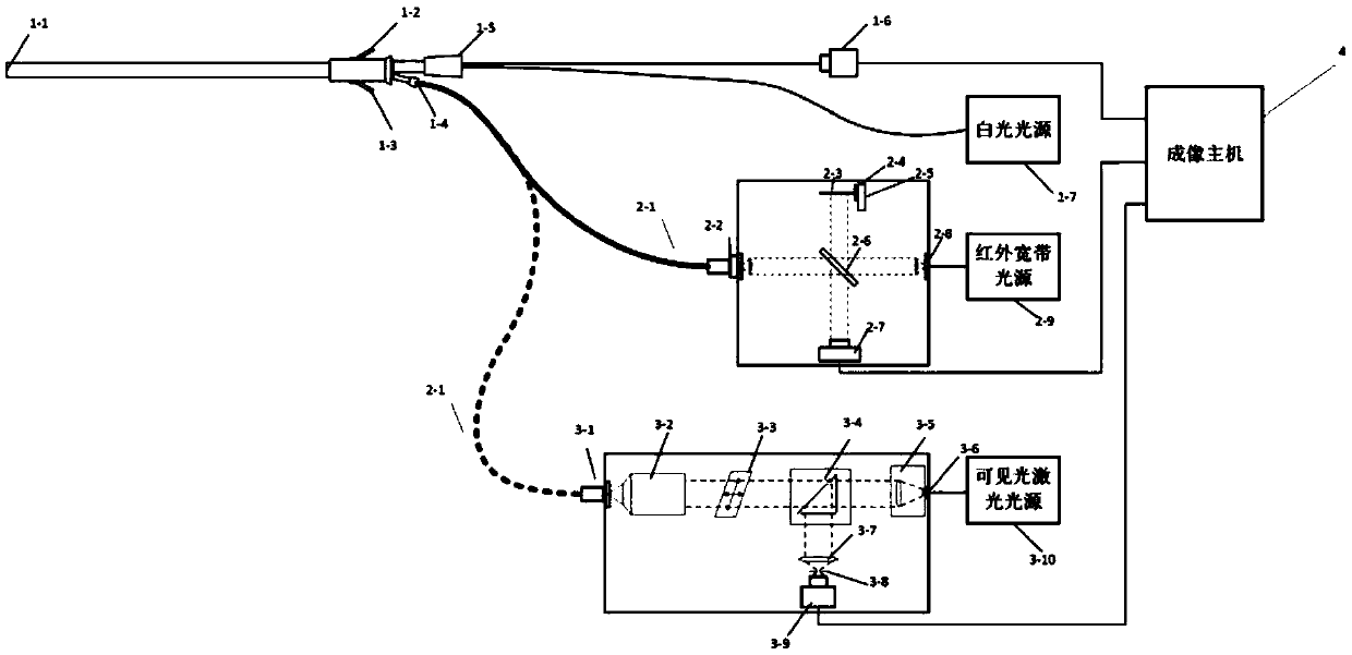
[0034] see figure 1 , which is a schematic diagram of an embodiment of the multi-mode hysteroscopy system of the present invention. As shown in the figure, the multi-mode hysteroscope system includes: a hysteroscope main mirror body, an optical coherence tomography imaging module, a confocal imaging module and an imaging host 4; wherein, the hysteroscope main mirror body includes an imaging probe 1-1, fluid inlet 1-2 and outlet 1-3 for perfusion, biopsy channel 1-4 for inserting tissue biopsy forceps, interface 1-5 for white light imaging, and imaging came...


PUM


Abstract

The invention discloses a multi-mode hysteroscope system and an obtaining method of the multi-mode hysteroscope system. The multi-mode hysteroscope system comprises a main hysteroscope body, an optical coherence tomography module, a co-focus imaging module and an imaging main unit. Compared with an existing hysteroscope, the multi-mode hysteroscope system has the advantage that ordinary white-light imaging, deep tomography and subcellsular level imaging can be achieved at the same time. The structural information of tissue within the depth range of 2 mm-3mm of the uterus epidermis can be provided through deep tomography based on the traditional white-light surface layer micro-imaging with the resolution ratio of 10 microns, early-stage hypodermic canceration can be detected, imaging with the high resolution ratio of 200 nm can be conducted on the epithelial layer of the mucosa through subcellsular level imaging, and canceration of a single cell can be detected accurately. According to the multi-mode hysteroscope system and the obtaining method of the multi-mode hysteroscope system, an important means is provided for early screening of metrocarcinoma, and the multi-mode hysteroscope system has broad application and market popularization prospects.

Description

technical field [0001] The invention relates to the technical field of endoscopes, in particular to a multi-mode hysteroscope system. Background technique [0002] Hysteroscopy is a new, minimally invasive gynecological diagnosis and treatment technology. It is a fiber light source endoscope used for inspection and treatment of uterine cavity. It is mainly used for diagnosis of symptoms: abnormal uterine bleeding, infertility or habitual Miscarriage, foreign bodies in the uterine cavity, submucosal myomectomy, endometrial polypectomy, intrauterine adhesions, mediastinal resection, and endometrial cancer. [0003] Cervicoscopy imaging system includes hysteroscope, energy system, light source system, perfusion system and imaging system; it uses the front part of the mirror body to enter the uterine cavity, uses white light for illumination, and the color high-definition has a magnifying effect on the observed part, so as to Intuitive and accurate, it has become the preferred ...

Claims


Application Information

Patent Timeline
Patent Type & Authority Patents(China)
IPC IPC(8): A61B1/00A61B1/05A61B5/00
CPCA61B1/00096A61B1/05A61B1/0661A61B5/0035A61B5/0066A61B5/0084A61B5/4325
Inventor 刘俊赵晖王翰林刘满林张浠安昕
Owner GUANGDONG OPTO MEDIC TECH CO LTD
Why Eureka
Try Eureka