Animal dorsal root ganglion-spinal cord fixed imaging and compression injury combining device

A technology of dorsal root ganglion and combined device, which is used in medical science, veterinary instruments, and musculoskeletal system evaluation, etc., can solve the problems of damage modeling, inability to complete, and inability to observe dorsal root ganglia.

Inactive Publication Date: 2015-07-15
GENERAL HOSPITAL OF PLA
View PDF9 Cites 4 Cited by
  • Summary
  • Abstract
  • Description
  • Claims
  • Application Information

AI Technical Summary

Problems solved by technology

However, it only provides a fixed observation device for the spinal cord, and cannot observe the

Method used


Image

  • Animal dorsal root ganglion-spinal cord fixed imaging and compression injury combining device
  • Animal dorsal root ganglion-spinal cord fixed imaging and compression injury combining device
  • Animal dorsal root ganglion-spinal cord fixed imaging and compression injury combining device

Examples


Embodiment Construction

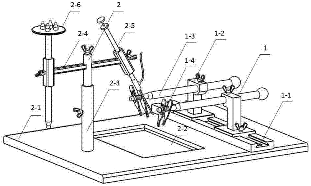
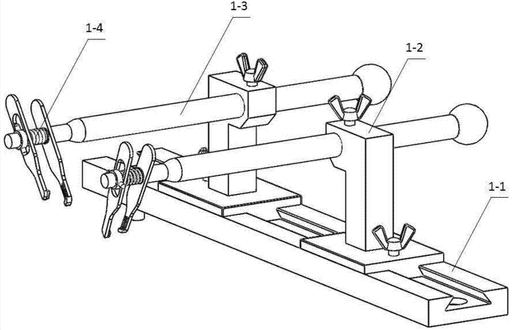
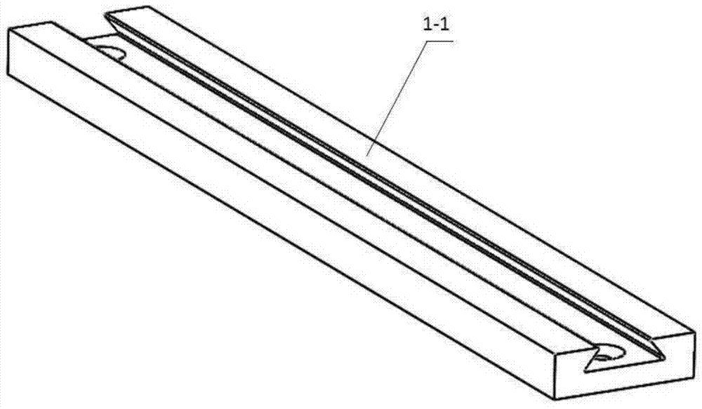
[0035] see specific Figure 1-11, the combined device of the present invention includes a dorsal root ganglion-spinal cord fixed imaging device 1 , and an integrated device 2 for microscopic fluorescent dye injection and spinal cord compression injury. The dorsal root ganglion-spinal cord fixation imaging device achieves clamping and fixation of single or multiple segments of the animal spine by adjusting the horizontal slider 1-2-1 and the L-shaped slider fixing block 1-2-2, Then perform single-stage or multi-stage laminectomy and pedicle resection, and lift the lateral sliding clip 1-4-1 of the spinal fixation clip 1-4 to rotate the spine in the medial direction so as to expose the lateral dorsal root ganglion. Purpose: After the operation, microinject the adeno-associated virus-green fluorescent protein and other dyes into the dorsal root ganglion through the microinjector 2-5-1 connected to the air pump connector 2-5-2 through the computer-controlled air pump device. Afte...


PUM


Abstract

The invention relates to an animal dorsal root ganglion-spinal cord fixed imaging and compression injury combining device, which comprises a dorsal root ganglion-spinal cord fixed imaging device and a microscopic fluorescent dye injection and spinal cord compression injury integrating device, wherein the animal dorsal root ganglion-spinal cord fixed imaging device (1) comprises a horizontal slide block guide rail (1-1), a fixing component (1-2), a slide rod (1-3) and a spine fixing clamp (1-4), and the microscopic fluorescent dye injection and spinal cord compression injury integrating device (2) comprises a vertical-rotating regulating side rod (2-3), a horizontal regulating component (2-4), a microscopic injection component (2-5) and a spinal cord compression component (2-6). The combining device has the advantages that multiple functions are integrated, the operation is simple and convenient, the switching can be realized at any time, different-degree spinal cord injury animal models can be conveniently manufactured, and after the fluorescent dye microscopic injection, a two-photon microscope is used for dynamically observing the pathological and physiological change process of the dorsal root ganglion-spinal cord injury in real time.

Description

technical field [0001] The invention discloses an animal dorsal root ganglion-spinal cord fixed imaging and compression injury device, which is mostly used in experimental research in the field of neurobiology. The device of the present invention can perform two-photon dynamic imaging of neurons and nerve fibers on single or multiple dorsal root ganglia and spinal cord segments in animals; and can form animal models of spinal cord injuries in different degrees. The device of the invention is of great value for studying the pathophysiological changes of dorsal root ganglion and spinal cord injury, and for establishing animal models of spinal cord compression injury. Background technique [0002] In the experimental research in the field of neurobiology, the traditional dorsal root ganglion and spinal cord imaging method is to make tissue slices. This technique requires the sacrifice of animals and removal of tissues, and tissue sectioning and staining after fixing the dorsal ...

Claims


Application Information

Patent Timeline
IPC IPC(8): A61B5/00A61D1/00
CPCA61B5/0059A61B5/45A61B2503/40A61D1/00A61D7/00
Inventor 张逸凌唐佩福张立海季欣然
Owner GENERAL HOSPITAL OF PLA
Why Eureka
Try Eureka