An intraoperative pathological tissue extractor

An extractor and tissue technology, applied in the direction of trocars, etc., can solve the problems of complicated operation process, easy to miss tissue, wrong diagnosis results, etc., to reduce the probability of bleeding, facilitate the collection process, and improve the accuracy.

Inactive Publication Date: 2017-09-26
FOURTH MILITARY MEDICAL UNIVERSITY
View PDF8 Cites 1 Cited by
  • Summary
  • Abstract
  • Description
  • Claims
  • Application Information

AI Technical Summary

Problems solved by technology

However, the disadvantages of this method are: 1. The operation process is complicated and difficult to master, and requires the support of equipment such as a cryostat; 2. Due to low temperature freezing during low temperature sectioning, the variability of tissues and cells is large, which affects the diagnosis to a certain extent The accuracy of the results; 3. The slices are relatively thick, the cells are large, and there are many layers, which is easy to cause wrong diagnostic results, but this inspection method has no room for extra thinking. Once an error is reported, it will cause serious and irreparable consequences; 4. More The most important thing is that this pathological detection method also takes 30 to 40 minutes to get the detection results, but the prolongation of the operation time will bring greater risks to the operation
The needle end of the syringe is beveled, but the diameter of some tissues is less than one centimeter. If the opening of the needle is too large, it is easy to miss the tissue or puncture the blood vessel, which makes the operation difficult; and a small amount of tissue cells extracted are easy to stay in the needle or attach to the needle. On the inner wall of the syringe, which brings difficulties to the subsequent collection process

Method used


Image

  • An intraoperative pathological tissue extractor
  • An intraoperative pathological tissue extractor
  • An intraoperative pathological tissue extractor

Examples


Embodiment Construction

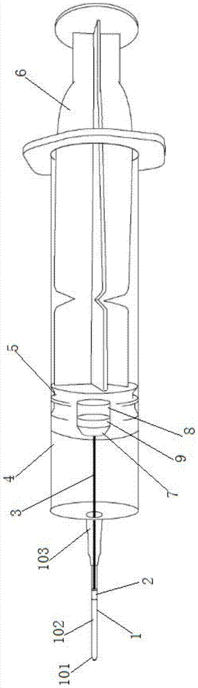
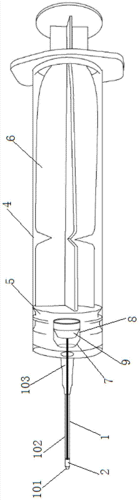
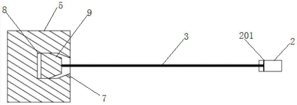
[0030] Cytological smear detection can quickly and effectively identify pathology during operation and provide an important basis for the formulation of operation plan. It has a high accuracy rate and can shorten the time of pathological detection to a few minutes, so it is a medical detection method with good application prospect. The most important point in cytology detection is to obtain pathological cell tissue with high quality as possible. At present, doctors use syringes instead of extraction devices to pierce organs and tissues to absorb a small amount of cells and tissues. However, syringes have too many disadvantages, mainly because the amount of tissue cells absorbed is small, and it is difficult to collect them quickly after extraction. The idea of ​​this scheme is to use another device to directly take out the extracted tissue cells on the basis of the syringe, so as to simplify the whole operation.

[0031] An intraoperative pathological tissue extractor, compri...


PUM


Abstract

The invention discloses an intraoperative pathological tissue extractor, which comprises a needle tube, a piston assembled in the needle tube, a push-pull rod for controlling the movement of the piston, and a needle head installed at the front end of the needle tube. The front end of the needle point of the needle head is circular The opening is equipped with an extraction cavity that can slide along the needle in the needle barrel of the needle, and the extraction cavity is connected with the piston through a connecting rod. The invention provides a special instrument capable of fast and convenient pathological extraction, which has simple structure, convenient operation and low cost; the circular needle end structure is designed to reduce the impact on the needle as much as possible without affecting the advance of the needle. The influence of other tissues can reduce the probability of bleeding and improve the accuracy of pathological cell collection; the setting of the connecting rod, extraction cavity and other position structures in this scheme enables all the tissues extracted at the end of the needle to enter the extraction cavity without Residing in the needle, the withdrawal chamber can be taken out directly from the back of the syringe, which greatly facilitates the tissue collection process.

Description

technical field [0001] The invention relates to a device for assisting in determining pathology, in particular to an extractor capable of rapidly collecting pathology during operation. Background technique [0002] Pathological diagnosis is the gold standard for clinically determining the surgical plan. Through the extraction and detection of pathological tissue, it can be determined whether the tumor is benign or malignant, which will directly affect the formulation of subsequent surgical plans. [0003] Traditional pathological tissue detection first needs to collect the tissue, and then use the fixative to fix the specimen and send it for inspection. This process takes one to two days and is often used in preoperative biopsy, but it is not applicable due to its long time In the determination of intraoperative pathology. [0004] Another inspection method that is often used is intraoperative rapid frozen section pathological examination. During the operation, the patholog...

Claims


Application Information

Patent Timeline
Patent Type & Authority Patents(China)
IPC IPC(8): A61B10/02
Inventor 李南林李永平樊菁王辉王廷张聚良李信杨玉庆魏洪亮
Owner FOURTH MILITARY MEDICAL UNIVERSITY
Why Eureka
Try Eureka