Animal spinal fixator

A fixation device and spine technology, applied in the field of fixation devices, can solve problems such as inability to realize observation, and achieve good braking effect

Inactive Publication Date: 2015-09-02
张金辉 +4
View PDF5 Cites 3 Cited by
  • Summary
  • Abstract
  • Description
  • Claims
  • Application Information

AI Technical Summary

Problems solved by technology

In the study of dorsal root ganglia, it is still in the in vitro test stage; in the study of spinal cord axis / dendrite, it is limited to the observation of the dorsal side of the animal under anesthesia, and cannot realize the observation in the awake state

Method used


Image

  • Animal spinal fixator
  • Animal spinal fixator
  • Animal spinal fixator

Examples


Embodiment 1

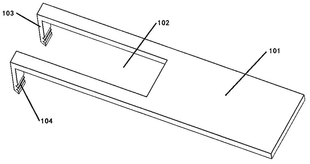
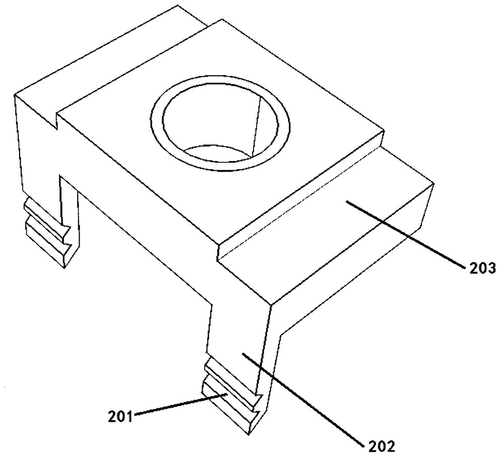
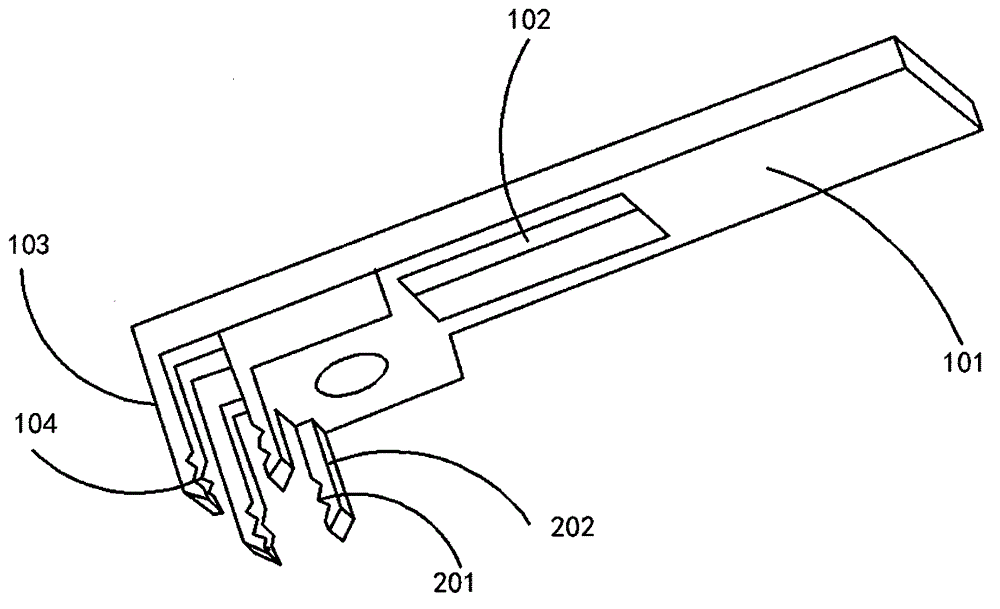
[0018] Such as figure 1 and figure 2 As shown, the present embodiment provides an animal spine fixation device, which includes a first clamp, a second clamp and an accommodating space; the first clamp is provided with a U-shaped groove 102; the front ends of both sides of the U-shaped groove 102 are bent to form two a first pinion 103; the second fixture cooperates with the U-shaped groove 102 and can slide in the U-shaped groove; the second fixture is provided with a second pinion 202 that cooperates with the first pinion; the accommodating space is connected with the two second pinches A prong 103 fits and communicates with the clamping portions of the first clamp and the second clamp.

[0019] Specifically, the hair on the back of the animal was removed and the skin was cut with a scalpel, part of the muscle tissue and tendons of the animal's spine were severed, and the first and second prongs were inserted into the muscles corresponding to the four transverse processes. ...

Embodiment 2

[0029] The first choice is to select adult transgenic fluorescent mice. Of course, fluorescent observation can also be achieved by injecting fluorescent agents into ordinary animals.

[0030] According to the body weight of the mice, 10 mg / mL of pentobarbital sodium was injected intraperitoneally, and the mice were injected with 0.08 mg of pentobarbital sodium and erythromycin eye ointment per gram of body weight.

[0031] After the mouse is anesthetized, transfer the mouse to a place far away from the operation area for shaving of the corresponding part, and clean it with 70% alcohol, then apply an appropriate amount of depilatory cream to the corresponding shaved area, and wait for 3-5 minutes after applying it evenly Scrape off the depilatory cream and rat hair with a scraper. Use a cotton ball dipped in normal saline to gently wipe off residual depilatory agent and rat hair, repeat this until it is clean, and finally dry the skin with a cotton ball.

[0032] Transfer the ...


PUM


Abstract

The invention relates to the field of fixators, in particular to an animal spinal fixator, comprising a first fixture, a second fixture and holding space. The first fixture is provided with a U-shaped channel; the front ends of the two sides of the U-shaped channel are bent, forming two first insert teeth; the second fixture fits the U-shaped channel and can slide in the same; the second fixture is provided with second insert teeth matching with the first insert teeth; the holding space fits the two first insert teeth and is communicated with clamping parts of the first and second fixtures. The animal spinal fixator allows an animal to be fixed and inspected after the animal wakens up; the animal can be fixed effectively; a ganglion and a spinal cord segment of a vertebral segment can be observed at the same time; in the research on spinal cord axon, the animal spinal fixator allows the spinal cord axon on the back of the animal to be observed.

Description

technical field [0001] The invention relates to the field of fixing devices, in particular to an animal vertebra fixing device. Background technique [0002] It is of unique value in neurophysiological and pathological research to observe the structure and function of the 1mm deep subcortex of animals under a nonlinear microscope in vivo or even in an awake state. In all current studies, it is a necessary condition for the study to ensure that the target position is completely immobilized during observation through surgery. In the study of the dorsal root ganglia, it is still in the in vitro test stage; in the study of the spinal cord axis / dendrite, it is limited to the observation of the dorsal side of the animal under anesthesia, and the observation under the awake state cannot be realized. Contents of the invention [0003] The object of the present invention is to provide an animal vertebra fixation device to solve the above problems. [0004] In order to achieve the...

Claims


Application Information

Patent Timeline
Patent Type & Authority Applications(China)
IPC IPC(8): A61D3/00
CPCA61D3/00
Inventor 张金辉
Owner 张金辉
Why Eureka
Try Eureka