Large-visual-field microscopic examination device and method for full-automatic immunohistochemistry

An immunohistochemistry, large field of view technology, applied in the field of medical equipment, can solve the problems of low detection efficiency and cumbersome detection process, and achieve the effect of improving detection efficiency and detection quality

Inactive Publication Date: 2015-11-25
苏州合惠生物科技有限公司
View PDF12 Cites 11 Cited by
  • Summary
  • Abstract
  • Description
  • Claims
  • Application Information

AI Technical Summary

Problems solved by technology

However, none of the fully automatic immunohistochemical instruments in the world can achieve true automation. After the sample is stained, it is taken under a microscope for microscopic examination. Medical personnel or professional operators are required t

Method used


Image

  • Large-visual-field microscopic examination device and method for full-automatic immunohistochemistry

Examples


Embodiment Construction

[0019] The present invention will be further described below in conjunction with the accompanying drawings.

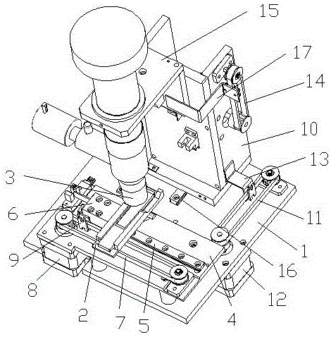
[0020] like figure 1 As shown, a large-field microscope inspection device for fully automatic immunohistochemistry includes a base 1, on which a slice stage 2 that can move horizontally relative to it is arranged, and above the base 1 is set There is a microscope lens 3 that can move horizontally and vertically relative to it, and the horizontal movement direction of the microscope lens 3 is perpendicular to the movement direction of the section stage 2, and a CCD camera is arranged above the microscope lens 3, Thus, the microscope lens 3 and the CCD camera can move back and forth, left and right, and up and down relative to the slice stage 2, which is convenient for focusing, and slice images can be collected through the movement of the microscope lens 3 and the CCD camera, which is convenient for later image processing and comparison. right.

[0021] In this embodi...


PUM


Abstract

The invention discloses a large-visual-field microscopic examination device for full-automatic immunohistochemistry. The large-visual-field microscopic examination device comprises a base. A section objective table capable of horizontally moving relative to the base is arranged on the base. A microscope lens capable of horizontally and longitudinally moving relative to the base is arranged above the base. The horizontal motion direction of the microscope lens is perpendicular to the moving direction of the section object table. A CCD camera is arranged above the microscope lens. The invention further discloses a large-visual-field microscopic examination method for full-automatic immunohistochemistry. The microscope lens and the CCD camera are arranged to be of the structure with the adjustable position, a complete section image is obtained by means of an image capture and splicing method, the section image and a database are compared to obtain a result, and full-automatic detection is achieved.

Description

technical field [0001] The invention relates to a large-field microscope inspection device and a detection method for automatic immunohistochemistry, belonging to the technical field of medical equipment. Background technique [0002] Immunohistochemistry is the gold standard for pathological detection and is widely used in tumor research and diagnosis. However, none of the fully automatic immunohistochemical instruments in the world can be truly fully automatic. After the sample is stained, it is taken under a microscope for microscopic examination. Medical personnel or professional operators are required to take out the sample and observe it under the microscope. , to judge the results, it cannot be interpreted by machine, and to realize full automation, not only the detection efficiency is relatively low, but also the detection process is relatively cumbersome. Contents of the invention [0003] The technical problem to be solved by the present invention is to provide ...

Claims


Application Information

Patent Timeline
IPC IPC(8): G01N21/84G01N35/00
Inventor 赵汗青丁代明
Owner 苏州合惠生物科技有限公司
Why Eureka
Try Eureka