Tumor grid organ model 3D printing method

A 3D printing and organ technology, applied in the medical field, can solve the problems of accurately judging the distance parameters of internal tumors and blood vessels in organs, high printing costs, and the inability to print organs themselves at the same time

Inactive Publication Date: 2016-01-13
王峻峰
View PDF8 Cites 9 Cited by
  • Summary
  • Abstract
  • Description
  • Claims
  • Application Information

AI Technical Summary

Problems solved by technology

The 3D printing model of ordinary tumors only prints the corresponding blood vessels and tumors, and the organs themselves are not printed. At the same time, only transparent silicone materials can be used for printing organs. The current printing cost is high, and tumors and organs cannot be directly measured through conventional 3D printing models. The distance between the surface and characteristic anatomical parts, and these data are helpful for clinical treatment such as surgery
If you want to print the organ itself and the blood vessels and tumors in the organ at the same time, you can ...

Method used


Image

  • Tumor grid organ model 3D printing method
  • Tumor grid organ model 3D printing method
  • Tumor grid organ model 3D printing method

Examples


Embodiment

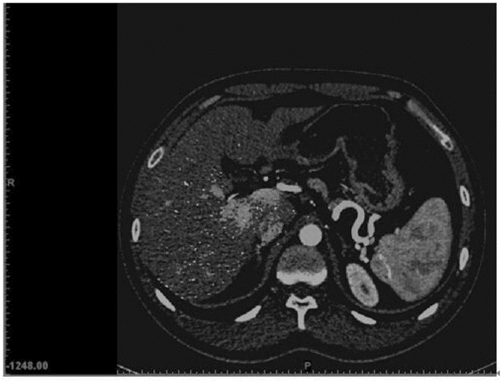
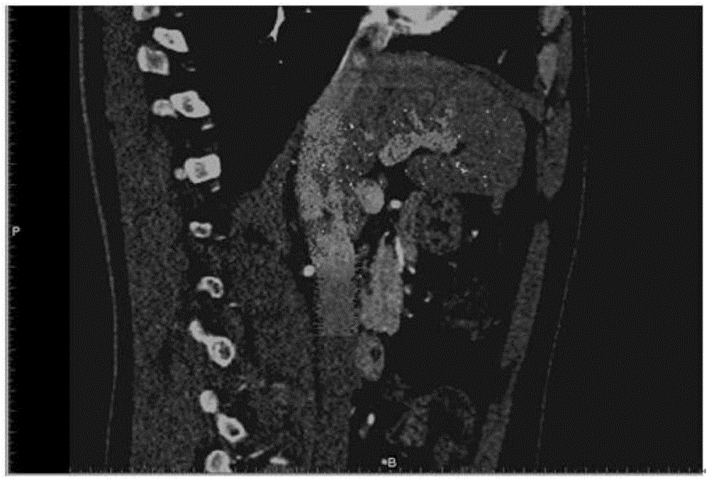
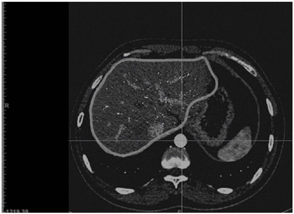
[0015] like Figure 1-5 As shown, the present invention provides a method for 3D printing a grid organ model of a tumor. Using the transverse section and sagittal plane of the human body in the supine position, the surface of the organ is used to establish a plurality of cross grids, and the grids are printed out. Organ models, and print tumors and blood vessels at the same time.

[0016] The original data (.dicom file) of medical imaging (CT or MRI), through special 3D modeling software, such as 3DSlicer, read the image and create 3D data, and finally print the generated 3D data through a "3D printer"; At the same time as the 3D data of blood vessels, the CT or MRI planes of the transverse and sagittal planes are selected to establish the 3D grid of the organ surface.

[0017] Manually outline the edge of the organ outline on the cross-section, and select a cylindrical line with a diameter of 3mm (conventional 3D modeling software can complete the line), and outline the edge...


PUM


Abstract

The invention relates to a tumor grid organ model 3D printing method. The method particularly comprises the following steps: a cross section and a sagittal section when a human body is in a supine position are used for building crossed multiple grids on the surface of an organ, while the 3D grids are built, 3D modeling software specially adopted for original data for medical imaging (CT or MRI) picks out an organ or a tumor or a blood vessel in need of 3D printing, and building of 3D data is carried out. 3D data for the organ or the tumor or the blood vessel are built at the same time, and organ surface 3D grid data are built. The 3D data are inputted to a 3D printer for 3D model printing. According to the printed model of the invention, the tumor can be intuitively observed via a hollow organ shell and the distance between the tumor and the organ surface can be measured, a guide is provided for surgical resection, and a spatial distance between the tumor or the blood vessel inside the organ and an anatomical site on the organ surface can be directly read from the model.

Description

technical field [0001] The invention belongs to the medical field and relates to 3D printing, in particular to a method for 3D printing a grid organ model of a tumor. Background technique [0002] The current 3D printing models of tumors and / or blood vessels mainly use ABS (ABS resin), PLA (polylactic acid) and PVA (polyvinyl alcohol), elastic silica gel, photosensitive resin, and the like. The 3D printing model of ordinary tumors only prints the corresponding blood vessels and tumors, and the organs themselves are not printed. At the same time, only transparent silicone materials can be used for printing organs. The current printing cost is high, and tumors and organs cannot be directly measured through conventional 3D printing models. The distance between the surface and characteristic anatomical parts, and these data are helpful for clinical treatment such as surgery. If you want to print the organ itself and the blood vessels and tumors in the organ at the same time, yo...

Claims


Application Information

Patent Timeline
IPC IPC(8): G09B23/30
CPCG09B23/303G09B23/30
Inventor 王峻峰司维
Owner 王峻峰
Why Eureka
Try Eureka