Method and device for X-ray image formation, scattering component calculation and reconstruction

An X-ray and image technology, applied in the field of medical images, can solve the problems of radiotherapy position error, reduced image accuracy, inability to achieve precise radiotherapy in the tumor area, etc., and achieve the effect of reducing the amount of scattering and good mechanical stability.

Active Publication Date: 2017-03-22
SHANGHAI UNITED IMAGING HEALTHCARE
View PDF4 Cites 0 Cited by
  • Summary
  • Abstract
  • Description
  • Claims
  • Application Information

AI Technical Summary

Problems solved by technology

In the process of radiotherapy, cone beam CT is used to obtain images of patients in real time and conduct image guidance for radiotherapy. The reduction of image accuracy leads to position errors of radiotherapy, which cannot achieve the goal of precise radiotherapy for tumor regions.

Method used


Image

  • Method and device for X-ray image formation, scattering component calculation and reconstruction
  • Method and device for X-ray image formation, scattering component calculation and reconstruction
  • Method and device for X-ray image formation, scattering component calculation and reconstruction

Examples


Embodiment Construction

[0025] In order to make the above objects, features and advantages of the present invention more comprehensible, specific implementations of the present invention will be described in detail below in conjunction with the accompanying drawings. In the following description, specific details are set forth in order to provide a thorough understanding of the present invention. However, the present invention can be implemented in many other ways than those described here, and those skilled in the art can make similar extensions without departing from the connotation of the present invention. Accordingly, the present invention is not limited to the specific embodiments disclosed below.

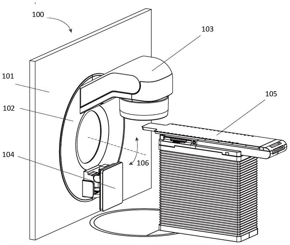
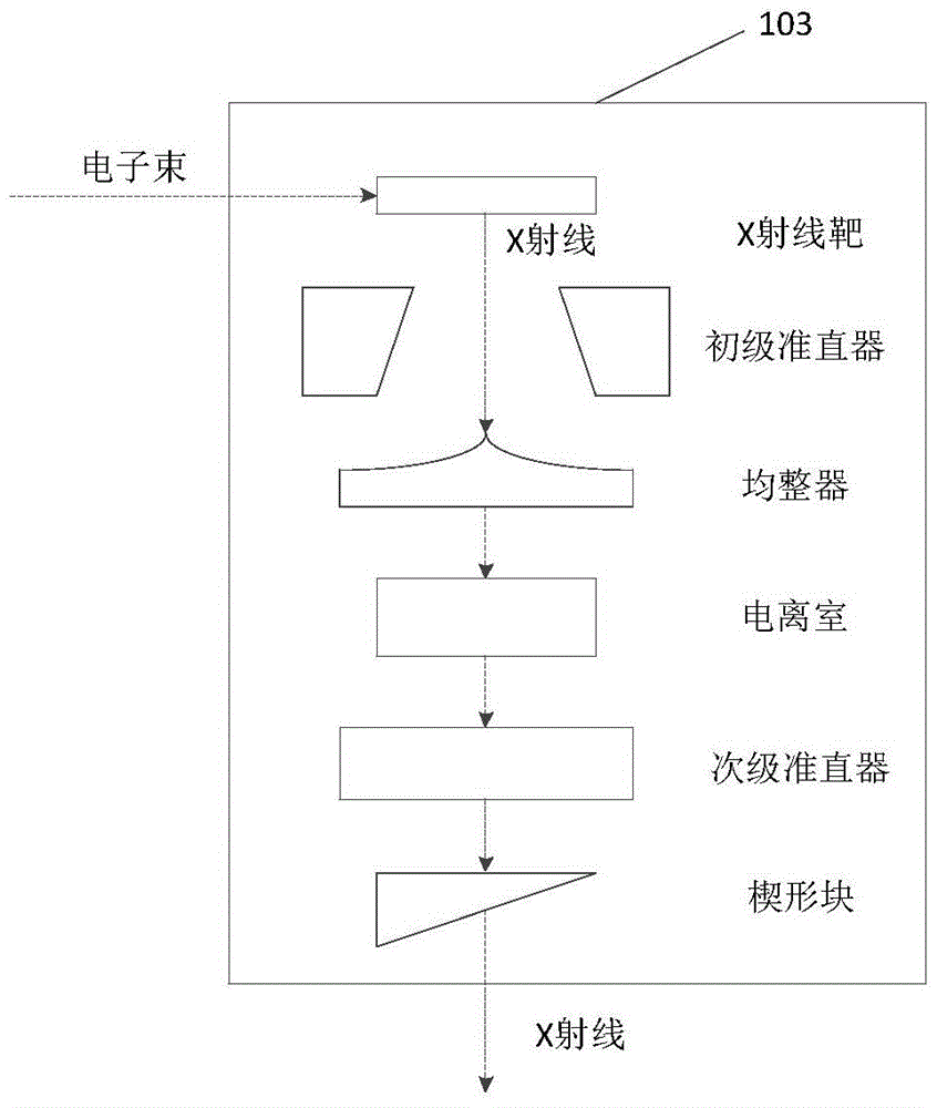
[0026] figure 1 is a structural diagram of a radiotherapy system, such as figure 1 As shown, the radiation therapy system 100 includes a fixed part 101 and a rotating part 102. The rotating part 102 is installed on the fixed part 101. The rotating part 101 can rotate around the central axis 106, s...


PUM


Abstract

The invention discloses a method and device for X-ray image imaging, X-ray image scattering component calculation, and X-ray image reconstruction. A radiographic image group; the collection is performed when the collimator moves along the first direction, and the X-ray image group is a group of X-ray images collected at different times of the collimator movement; extracting the sub-region images of each X-ray image in the X-ray image group; combining the sub-region images along the first direction to form a modulated X-ray image. The technical solution of the present invention realizes the flexible modulation of the X-ray intensity distribution by using the collimator, without adding additional hardware, and calculates the scattering components in the X-ray image, finally reducing the scattering amount of the X-ray image.

Description

technical field [0001] The invention relates to the field of medical images, in particular to a method and device for forming, calculating and reconstructing X-ray images. Background technique [0002] In X-ray images, scattered rays have always been an important factor leading to unsatisfactory image quality. Especially for X-ray systems with a larger detector area, such as radiotherapy equipment, the cone beam computed tomography (cone beam computed tomography) used uses the Electronic Portal Imaging Device (EPID, Electronic Portal Imaging Device) as the receiving X-ray system. X-ray detectors, scattered rays account for a large proportion of the X-rays they receive. Scattered rays cause cupping artifacts on the image, reduce the contrast of the image, and greatly reduce the image accuracy of the reconstructed image. During radiation therapy, cone-beam CT is used to obtain images of patients in real time and image-guid radiation therapy. The reduction of image accuracy l...

Claims


Application Information

Patent Timeline
Patent Type & Authority Patents(China)
IPC IPC(8): A61B6/00
CPCA61B6/03A61B6/06A61B6/4035A61B6/5205A61B6/54G21K1/043A61N2005/1054A61B6/5282A61N5/1045A61N5/1049A61N2005/1095G06T2207/10116G06T5/002G06T5/003G06T5/50G06T2207/20212G06T5/001G06T2207/20224A61B6/032A61B6/5217
Inventor 李贵杨宏成徐璐
Owner SHANGHAI UNITED IMAGING HEALTHCARE
Why Eureka
Try Eureka