Medical image organ recognition method and segmentation method

A medical image and recognition method technology, which is applied in the field of organ recognition and segmentation in medical images, can solve the problems of repeated calculation and large amount of calculation, recognition errors, and a large number of Haar features, and achieve strong self-adaptability and removal Boundary noise, the effect of improving recognition efficiency

Active Publication Date: 2016-03-09
SHANGHAI UNITED IMAGING HEALTHCARE
View PDF6 Cites 46 Cited by
  • Summary
  • Abstract
  • Description
  • Claims
  • Application Information

AI Technical Summary

Problems solved by technology

[0003] Manually identifying and marking the body part where the current CT image is located by the doctor requires a lot of repetitive work by the doctor, and the efficiency is low
The existing methods for automatic recognition of body parts in CT images can be mainly divided into three types: (1) body part recognition based on the header information of Digital Imaging and Communications in Medicine (DICOM) [1], usually the DICOM file header contains CT images Scanned label information, but due to differences in various cultures and languages, labels recorded in different languages ​​will increase the difficulty of accurately identifying DICOM file header information, and even wrong DICOM file header information will lead to recognition errors; (2) Based on gray value features The method is mainly based on the different degrees of attenuation of X-rays when passing through different tissue components of the body. The gray value distribution of different tissue components in the CT image is different. The method based on gray value features is based on the main tissue components of the body in CT. The prior knowledge of the gray value distribution in the image can be used to divide the body parts. However, this method has a low recognition rate for the head and pelvis [2]; (3) The method based on machine learning is mainly divided into training and There are two stages of testing. In the training stage, the Haar image features corresponding to the key organs of the body are extracted, and a large number of positive and negative samples are constructed. By training the AdaBoost classifier, the effective Haar feature sequences corresponding to the organs and their corresponding weights are extracted. In the testing stage, the input to be The measured image, calculate the Haar feature value of the image, compare it with the existing training results, and determine whether the image is a positive sample [3]. Sampling or downsampling, and the number of Haar features used is large, there are problems of repeated calculations, large calculations, and low operational efficiency

Method used


Image

  • Medical image organ recognition method and segmentation method
  • Medical image organ recognition method and segmentation method
  • Medical image organ recognition method and segmentation method

Examples


Embodiment Construction

[0046] In order to make the above objects, features and advantages of the present invention more obvious and comprehensible, specific implementations of the present invention will be described in detail below in conjunction with the accompanying drawings and embodiments.

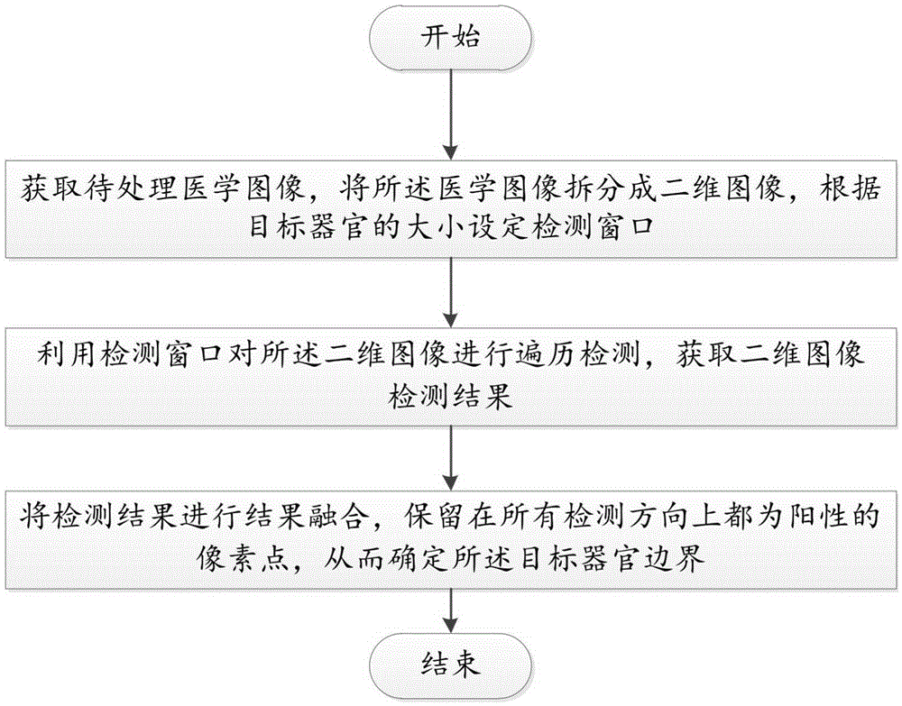
[0047] In clinical diagnosis, medical images play an important role. Medical image segmentation is the first stage of medical image data analysis and visualization. The first prerequisites and key steps. Accurately judging the position of human body organs in medical images before medical image segmentation plays an important role in improving the accuracy of segmentation. Such as figure 1 Shown is a flow chart of the method for identifying organs in medical images according to the present invention, which mainly includes the following steps:

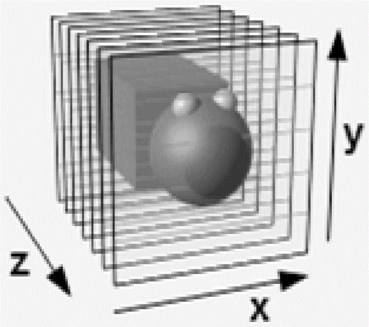
[0048] S10. Acquire a medical image to be processed, split the medical image into several two-dimensional images in X, Y, and Z axis directions, and set a detection...


PUM


Abstract

The invention discloses a medical image organ recognition method. The method comprises steps: a to-be-processed medical image is acquired, the medical image is segmented into a plurality of two-dimensional images in X-axis, Y-axis and Z-axis directions respectively, and a detection window is set according to the size of a target organ; the detection window is used to carry out traversing detection on the two-dimensional images respectively according to a set detection step length, and detection results in the X-axis, Y-axis and Z-axis directions are acquired; and result fusion is carried out on the detection results, pixel points which are detected to be positive in the X-axis, Y-axis and Z-axis directions are kept, and a target organ boundary is determined. The medical image organ recognition method of the invention can quickly and accurately recognize the target organ area, the target organ boundary is determined and the adaptive ability is strong. In addition, the invention also provides a medical image organ segmentation method.

Description

【Technical field】 [0001] The invention relates to the field of medical image processing, in particular to an identification method and a segmentation method of organs in medical images. 【Background technique】 [0002] With the increasing maturity of medical imaging technology and the wide application of various medical imaging equipment in hospitals, information images of human internal tissues can be obtained conveniently and non-destructively, and these information can be effectively processed by image processing technology to assist doctors in diagnosis. Even surgical planning, etc., has significant social benefits and broad application prospects. For example, a computed tomography (CT) image is a matrix composed of a certain number of pixels arranged in different gray levels from black to white. The pixels can reflect the X-ray absorption coefficient of the corresponding voxel, while different gray levels reflect the organs or organs. The degree to which tissues absorb ...

Claims


Application Information

Patent Timeline
Patent Type & Authority Applications(China)
IPC IPC(8): G06T7/00
CPCG06T7/0012G06T2207/10072G06T2207/10081G06T2207/10088G06T2207/10104G06T2207/10108G06T2207/30004G06T2207/30008
Inventor 田野李强
Owner SHANGHAI UNITED IMAGING HEALTHCARE
Why Eureka
Try Eureka