Method for automatically identifying liver tumor type in ultrasonic image

An ultrasonic imaging and automatic recognition technology, applied in character and pattern recognition, ultrasonic/sonic/infrasonic diagnosis, image enhancement, etc., can solve the problem of no type of lesion identification, no identification of focal liver lesions, etc., to overcome the lesion area Effect

Inactive Publication Date: 2016-03-30
SUN YAT SEN UNIV
View PDF0 Cites 33 Cited by
  • Summary
  • Abstract
  • Description
  • Claims
  • Application Information

AI Technical Summary

Problems solved by technology

Determining the location of the lesion is a very important step in the identification of this method, but the above metho

Method used


Image

  • Method for automatically identifying liver tumor type in ultrasonic image
  • Method for automatically identifying liver tumor type in ultrasonic image
  • Method for automatically identifying liver tumor type in ultrasonic image

Examples


Embodiment Construction

[0044] The present invention will be further described below in conjunction with the drawings, but the embodiments of the present invention are not limited to this.

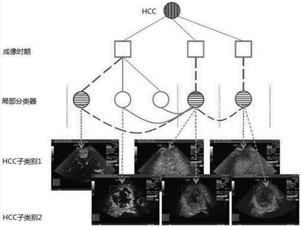
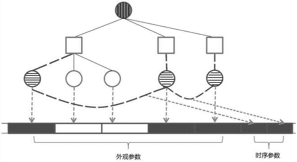
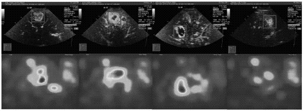
[0045] The present invention mainly provides a method for automatically identifying liver tumor types in ultrasound images, which includes two parts: a method of expressing liver tumors in ultrasound images; and automatically determining the location, time and size of the lesion in ultrasound images, and Method of performing lesion identification.

[0046] To facilitate the description, define the following key terms:

[0047] A "vector" is a set of numbers arranged in sequence, which can be represented by a computer programming language, such as an array in the C language.

[0048] "Model" is a set of rules that can take the ultrasound image video of a case as input and output as a possible value whether it belongs to a certain type of lesion. In this method, the model divides the input into a vector with a certain str...


PUM


Abstract

The invention relates to the field of medical image processing, in particular to a method for automatically identifying a liver tumor type in an ultrasonic image. The method can provide auxiliary diagnosis when a doctor diagnoses a liver lesion according to a CEUS medical image, wherein the auxiliary diagnosis comprises the process of giving out a lesion identification result, the time when the lesion is relatively remarkable and a position where the lesion is relatively remarkable. The method specifically comprises: representing a CEUS image with a plurality of regions of interest (ROI); distinguishing different lesions by expressions and changes of the ROI in time and space; and representing a space-time relationship among the ROI by establishing models in time and space at the same time, wherein the models can determine relatively proper ROI and related parameters of the models according to existing CEUS lesion samples with an iterative learning method. After a sample is given out, the proper ROI can be determined by removing part of improper ROI in advance and a fast search method, and reference diagnosis is given out for the lesion.

Description

Technical field [0001] The present invention relates to the field of medical image processing, and more specifically, to a method for automatically identifying liver tumor types in ultrasound images. Background technique [0002] Liver tumors are the fifth most common tumor and the second leading cause of death in cancer. Focal liver lesions (FLLs) are abnormal solid or cystic masses in the liver. The early detection and diagnosis of FLLs in liver cancer are of great significance to the treatment of liver cancer. In the process of diagnosis, medical imaging plays a very important role. Especially in recent years, with the development of imaging technology, medical imaging has become more and more important in diagnosis. Medical imaging includes electronic computed tomography (CT), magnetic resonance imaging (MRI), ultrasound imaging (US), etc., among which CT and MRI require high costs, complicated equipment, and CT imaging can also cause ionizing radiation. Ultrasound imaging ...

Claims


Application Information

Patent Timeline
IPC IPC(8): G06T7/00G06K9/62G06V10/25
CPCG06T7/0016G06T2207/20104G06T2207/30056G06T2207/10132G06F18/214A61B8/481A61B8/085A61B8/469A61B8/5223G06T7/0014G06T2207/20076G06T2207/20081G06T2207/30096G16H50/30G06V10/25G06V10/87G06F18/285G06V2201/03G06F18/251G06F18/2148G06F18/24147G06T7/62G06T7/70G06T2207/20072
Inventor 林倞曹擎星王青江波
Owner SUN YAT SEN UNIV
Why Eureka
Try Eureka