Method for induced differentiation of human amniotic mesenchyme cells in vitro into insulin-secreting cells

An amniotic mesenchyme, insulin secretion technology, applied in the biological field, can solve the problems of not expressing telomerase gene, not having long-term self-renewal and the ability to generate single cell clones, and not being tumorigenic.

Inactive Publication Date: 2016-10-12
吴卫江
View PDF0 Cites 5 Cited by
  • Summary
  • Abstract
  • Description
  • Claims
  • Application Information

AI Technical Summary

Problems solved by technology

Amniotic epithelial cells (AECs) have the potential to differentiate into three germ layer cells, but AECS do not express the telomerase gene, so they cannot be call

Method used


Image

  • Method for induced differentiation of human amniotic mesenchyme cells in vitro into insulin-secreting cells

Examples


Embodiment Construction

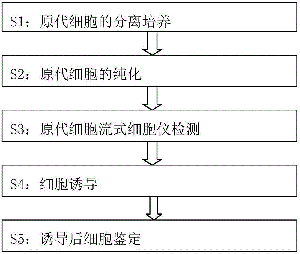
[0013] The method for directional differentiation of human amniotic mesenchymal cells into insulin-secreting cells in vitro comprises the following steps:

[0014] S1: Isolation and culture of primary cells: Under aseptic conditions, take fresh placenta discarded after delivery (with the consent of the family members), bluntly separate the amniotic membrane from the chorionic villi, and place it in a placenta containing double antibodies (100U / ML penicillin, 100UG / ML streptomycin). prime) D-HANKS solution for repeated washing. Cut the rinsed amniotic membrane into about 1*1mm pieces with ophthalmic scissors, add 2.5G / L trypsin to digest at 37°C for 10 minutes, add RPMI1640 medium containing 5% calf serum to stop the digestion, and gently blow and mix for 200 Filter with a mesh cell sieve, add 1.0g / L type Ⅱ collagenase digestion solution to the filtered amnion tissue, digest at 37°C for 0.5h, stop digestion again with 5% calf serum RPMI1640, gently blow and mix, and then use a ...


PUM

PropertyMeasurementUnit
Thicknessaaaaaaaaaa

Abstract

The invention provides a method for induced differentiation of human amniotic mesenchyme cells in vitro into insulin-secreting cells. The steps include: isolation culture of primary cells; purification of primary cells; primary cell identification by a flow cytometer; 2-mercaptoethanol, B27, bFGF, and nicotinamide induction; and cell morphology and function identification after induction. The method for induced differentiation of human amniotic mesenchyme cells in vitro into insulin-secreting cells provided by the invention induces human amniotic mesenchymal cells to differentiate into nestin positive cells, induces nestin positive cell amplification and differentiation into precursor cells, and induces insulin-secreting cell maturation, confirms that amniotic mesenchyme cells have the possibility of transformation into insulin-secreting cells in specific environment, amniotic mesenchyme cells have wide sources, and no ethical obstacle exists, therefore the method provides a new idea for clinical stem cell transplantation.

Description

technical field [0001] The invention relates to the field of biotechnology, in particular to a method for directional differentiation of human amniotic mesenchymal cells into insulin-secreting cells in vitro. Background technique [0002] The amniotic membrane is located on the surface of the fetal chorion and is a smooth, avascular, nerveless, and lymphatic transparent film. Amnion tissue is mainly composed of two types of cells: amniotic epithelial cells (AECs) in the ectoderm and amniotic mesenchyme cells (AMCs) in the mesoderm. Amniotic epithelial cells (AECs) have the potential to differentiate into three germ layer cells, but AECS do not express the telomerase gene, so they cannot be called stem cells in strict definition, and do not have the ability of long-term self-renewal and generation of single cell clones. Not tumorigenic (Toshio, et al, 2005). Amnion epithelial cells are easy to obtain and have abundant sources. Their pluripotent stem cell nature and low immu...

Claims


Application Information

Patent Timeline
IPC IPC(8): C12N5/071C12N5/077
Inventor 吴卫江
Owner 吴卫江
Why Eureka
Try Eureka